. 2016 Dec;46(6):296-298.
doi: 10.4274/tjo.24445. Epub 2016 Dec 1.

Focal Choroidal Excavation

Affiliations

Focal Choroidal Excavation

Zafer Cebeci et al. Turk J Ophthalmol. 2016 Dec.

Abstract

Focal choroidal excavation is a choroidal pit that can be detected by optical coherence tomography. Central serous chorioretinopathy, choroidal neovascularization and polypoidal choroidal vasculopathy are pathologies associated with focal choroidal excavation. In this article, we present the follow-up and treatment outcomes of three eyes of two patients with focal choroidal excavation.

Keywords: central serous chorioretinopathy; choroidal neovascularization; optical coherence tomography.


Conflict of interest statement

No conflict of interest was declared by the authors. Financial Disclosure: The authors declared that this study received no financial support.

Figures
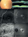
Figure 1
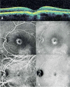
Figure 1. Case 1 before treatment: left eye fundus photography showing macular pigment epithelium changes (a), right eye optic coherence tomography showing focal choroidal excavation (b), left eye optic coherence tomography showing subfoveal choroidal excavation and subretinal fluid (c), left eye fundus fluorescein angiography showing early stage hyperfluorescence as a window defect (d), left eye fundus fluorescein angiography showing intensified hyperfluorescence in the late phase (e), left eye indocyanine green angiography showing choroidal vessel dilation in the early phase (f), left eye indocyanine green angiography showing late phase hyperfluorescence due to leakage from choroidal vessels (g)
Figure 2
Figure 2. Case 1 at 18 months after photodynamic therapy: focal choroidal excavation and subretinal fluid do not appear on left eye optic coherence tomography (a), left eye fundus fluorescein angiography, early phase (b), left eye fundus fluorescein angiography showing late phase hyperfluorescence due to a window defect (c) left eye indocyanine green angiography showing resolution of choroidal vessel dilation in the early phase (d), late phase hyperfluorescence is no longer apparent on left eye indocyanine green angiography
Figure 3
Figure 3. Case 2 pre- and post-treatment appearance: left eye fundus photography showing macular pigment epithelium changes (a), left eye fundus fluorescein angiography, early phase (b), left eye fundus fluorescein angiography showing hyperfluorescence intensifying in the late phase (c), left eye optic coherence tomography showing focal choroidal excavation and an overlying hyporeflective area (d), left eye fundus photograph at 2 years post-treatment showing pigment epithelium changes at the macula (e), left eye fundus fluorescein angiography, early phase at 2 years post-treatment (f), left eye fundus fluorescein angiography at 2 years post-treatment showing hyperfluorescence slightly intensified in the late phase (g), left eye optic coherence tomography at 2 years post-treatment showing conforming excavation and the absence of a hyporeflective area (h)

References

    1. Lee CS, Woo SJ, Kim YK, Hwang DJ, Kang HM, Kim H, Lee SC. Clinical and spectral-domain optical coherence tomography findings in patients with focal choroidal excavation. Ophthalmology. 2014;121:1029–1035. - PubMed
    1. Jampol LM, Shankle J, Schroeder R, Tornambe P, Spaide RF, Hee MR. Diagnostic and therapeutic challenges. Retina. 2006;26:1072–1076. - PubMed
    1. Margolis R, Mukkamala SK, Jampol LM, Spaide RF, Ober MD, Sorenson JA, Gentile RC, Miller JA, Sherman J, Freund KB. The expanded spectrum of focal choroidal excavation. Arch Ophthalmol. 2011;129:1320–1325. - PubMed
    1. Obata R, Takahashi H, Ueta T, Yuda K, Kure K, Yanagi Y. Tomographic and angiographic characteristics of eyes with macular focal choroidal excavation. Retina. 2013;33:1201–1210. - PubMed
    1. Kobayashi W, Abe T, Tamai H, Nakazawa T. Choroidal excavation with polypoidal choroidal vasculopathy: a case report. Clin Ophthalmol. 2012;6:1373–1376. - PMC - PubMed

LinkOut - more resources