

Foot and ankle fractures during childhood: review of the literature and scientific evidence for appropriate treatment
- PMID: 28050532
- PMCID: PMC5198067
- DOI: 10.1016/j.rboe.2016.09.001
Foot and ankle fractures during childhood: review of the literature and scientific evidence for appropriate treatment
Abstract
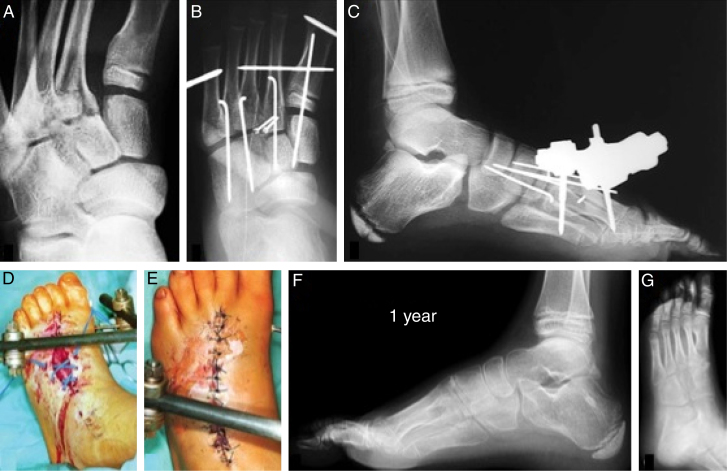
Foot and ankle fractures represent 12% of all pediatric fractures. Malleolar fractures are the most frequent injuries of the lower limbs. Hindfoot and midfoot fractures are rare, but inadequate treatment for these fractures may results in compartment syndrome, three-dimensional deformities, avascular necrosis and early post-traumatic arthritis, which have a significant impact on overall foot and ankle function. Therefore, the challenges in treating these injuries in children are to achieve adequate diagnosis and precise treatment, while avoiding complications. The objective of the treatment is to restore normal anatomy and the correct articular relationship between the bones in this region. Moreover, the treatment needs to be planned according to articular involvement, lower-limb alignment, ligament stability and age. This article provides a review on this topic and presents the scientific evidence for appropriate treatment of these lesions.
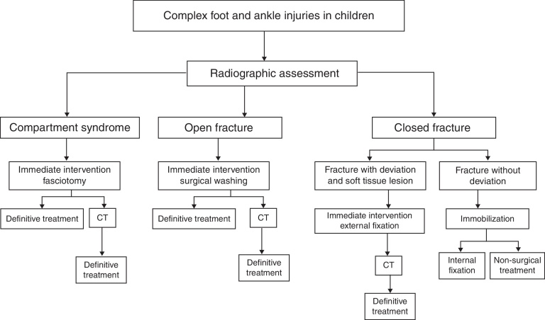
As fraturas do tornozelo e do pé representam 12% de todas as fraturas pediátricas. Fraturas maleolares são as lesões mais frequentes dos membros inferiores; fraturas do retropé e mediopé são raras, mas o seu tratamento inadequado pode resultar em síndrome de compartimento, deformidades tridimensionais, necrose avascular e osteoartrose pós-traumática precoce, as quais apresentam impacto significativo na função global do tornozelo e pé. Portanto, os desafios no tratamento dessas lesões na criança são o diagnóstico adequado e tratamento preciso para se evitarem as complicações. O objetivo do tratamento é restaurar a anatomia normal e a relação articular correta entre os ossos da região. Além disso, o tratamento deve ser planejado de acordo com acometimento articular, o alinhamento dos membros inferiores, a estabilidade ligamentar e a idade. O algoritmo de tratamento dos traumas complexos do tornozelo e pé na infância é descrito. Este artigo apresenta uma revisão sobre o tema e as evidências científicas para o tratamento adequado dessas lesões.
Keywords: Ankle joint; Calcaneus; Child; Talus.
Figures

References
-
- Crawford A.H., Al-Sayyad M.J. Fractures and dislocations of the foot and ankle. In: Green N.E., Swiontkowski M.F., editors. Skeletal trauma in children. Saunders; Philadelphia: 2003. pp. 516–537.
-
- Petit C.J., Lee B.M., Kasser J.R., Kocher M.S. Operative treatment of intraarticular calcaneal fractures in the pediatric population. J Pediatr Orthop. 2007;27(8):856–862. - PubMed
-
- Rammelt S., Schneiders W., Fitze G., Zwipp H. Foot and ankle fractures in children. Orthopade. 2013;42(1):45–54. - PubMed
-
- Lauge-Hansen N. Fractures of the ankle. II. Combined experimental–surgical and experimental–roentgenologic investigations. Arch Surg. 1950;60(5):957–985. - PubMed
-
- Dias L.S., Tachdjian M.O. Physeal injuries of the ankle in children: classification. Clin Orthop Relat Res. 1978;(136):230–233. - PubMed
Publication types
LinkOut - more resources
Full Text Sources
Other Literature Sources