

Ultrasonography has a diagnostic value in the assessment of cervical radiculopathy: A prospective pilot study
- PMID: 28050690
- PMCID: PMC5491566
- DOI: 10.1007/s00330-016-4704-9
Ultrasonography has a diagnostic value in the assessment of cervical radiculopathy: A prospective pilot study
Abstract
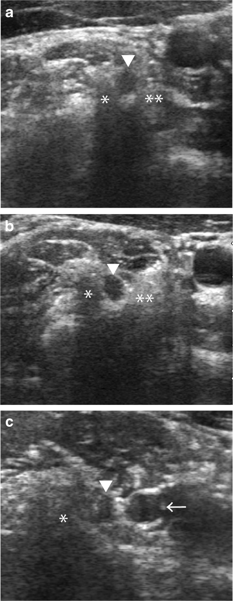
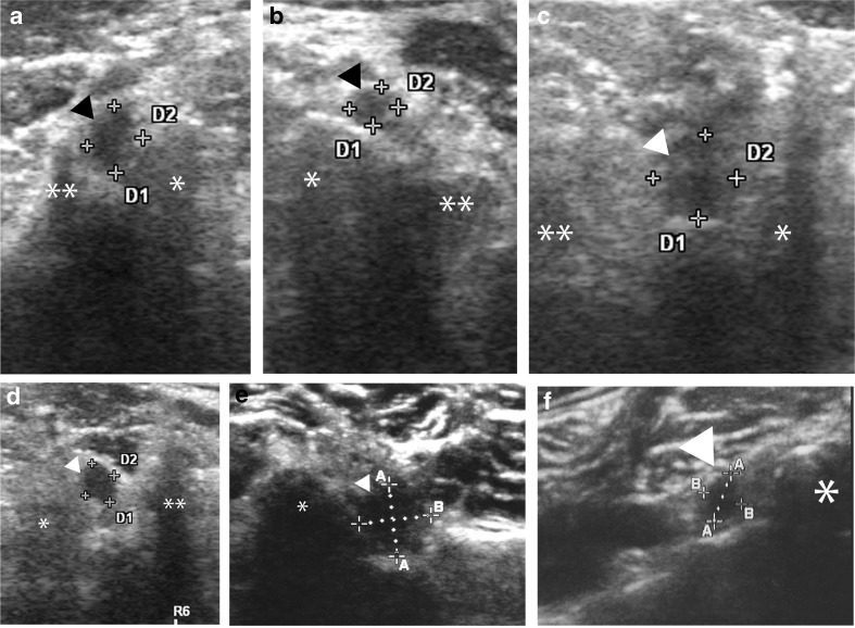
Objective: This study investigated the diagnostic accuracy of the difference in the cross-sectional areas (CSAs) of affected cervical nerve roots (NRs) for diagnosing cervical radiculopathy (CR).
Methods: In total, 102 CR patients and 219 healthy volunteers were examined with ultrasound. The CSA of the cervical NR at each level was measured on the affected side and the contralateral side in CR patients by blinded ultrasonographic technicians. The difference between the CSAs of CR patients and normal volunteers and the difference in the laterality of CSA at the same affected level (ΔCSA) were calculated for each cervical level.
Results: The CSAs of the affected NRs in CR patients were significantly larger than those of the unaffected NRs in CR patients and those of the control group at the C5, C6 and C7 levels (P<0.005). ΔCSA was also significantly larger in the CR group at all levels (P<0.001). A receiver operating characteristic analysis demonstrated that the threshold values were 9.6 mm2 (CSA) for C5NR and 15 mm2 for both C6NR and C7NR.
Conclusions: This study revealed that the CSAs of affected NRs were enlarged and that the laterality of the CSA (ΔCSA) was greater in CR patients than in control patients.
Key points: • Cervical radiculopathy is diagnosed through ultrasonographic measurement of the CSAs. • The CSAs of affected nerve roots were significantly enlarged. • The ΔCSA in the CR group was significantly higher than in the control group. • Diagnostic CSA and ΔCSA thresholds were identified.
Keywords: Cervical nerve root; Cervical radiculopathy; Cross-sectional area; Ultrasonography; Ultrasound.
Figures
References
MeSH terms
LinkOut - more resources
Full Text Sources
Other Literature Sources
Medical
Miscellaneous
