. 2017 Feb;25(3):315-323.
doi: 10.1038/ejhg.2016.183. Epub 2017 Jan 4.

Severe neurodegenerative disease in brothers with homozygous mutation in POLR1A

Affiliations

Severe neurodegenerative disease in brothers with homozygous mutation in POLR1A

Bülent Kara et al. Eur J Hum Genet. 2017 Feb.

Abstract

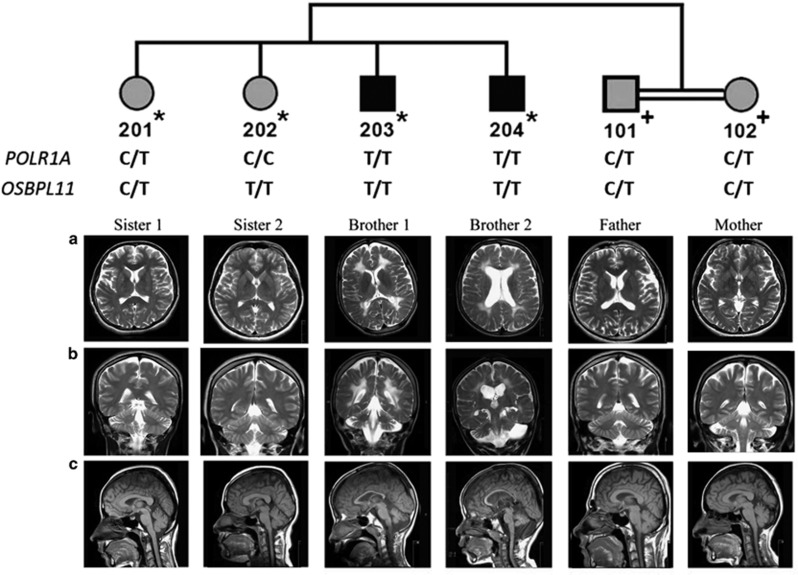
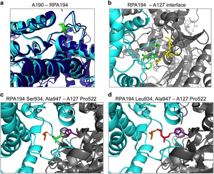
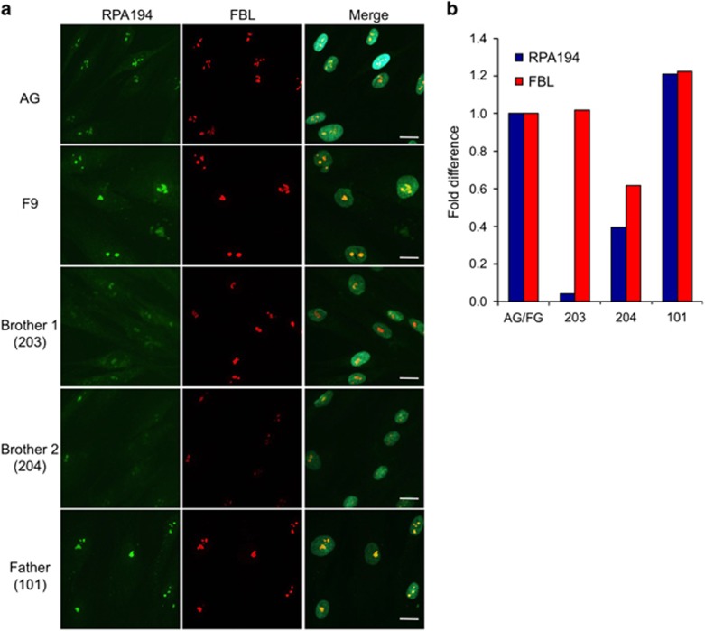
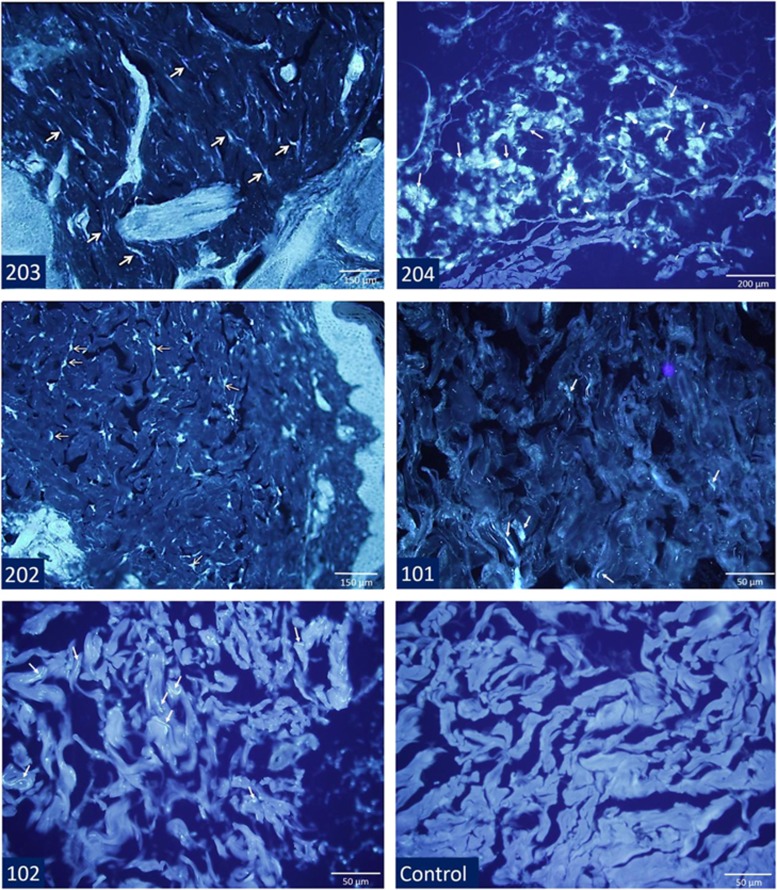
In two brothers born to consanguineous parents, we identified an unusual neurological disease that manifested with ataxia, psychomotor retardation, cerebellar and cerebral atrophy, and leukodystrophy. Via linkage analysis and exome sequencing, we identified homozygous c.2801C>T (p.(Ser934Leu)) in POLR1A (encoding RPA194, largest subunit of RNA polymerase I) and c.511C>T (p.(Arg171Trp)) in OSBPL11 (encoding oxysterol-binding protein-like protein 11). Although in silico analysis, histopathologic evidence and functional verification indicated that both variants were deleterious, segregation with the patient phenotype established that the POLR1A defect underlies the disease, as a clinically unaffected sister also was homozygous for the OSBPL11 variant. Decreased nucleolar RPA194 was observed in the skin fibroblasts of only the affected brothers, whereas intracellular cholesterol accumulation was observed in the skin biopsies of the patients and the sister homozygous for the OSBPL11 variant. Our findings provide the first report showing a complex leukodystrophy associated with POLR1A. Variants in three other RNA polymerase subunits, POLR1C, POLR3A and POLR3B, are known to cause recessive leukodystrophy similar to the disease afflicting the present family but with a later onset. Of those, POLR1C is also implicated in a mandibulofacial dysostosis syndrome without leukodystrophy as POLR1A is. This syndrome is absent in the family we present.


Conflict of interest statement

The authors declare no conflict of interest.

Figures

Figure 1
Figure 1
The pedigree and cranial MR images. DNA samples subjected to SNP genotyping are indicated by * on the pedigree, and those for variant testing are indicated by +. POLR1A and OSBPL11 variant genotypes are given. MR images of the brothers revealed enlarged ventricles, cortical sulci and subarachnoid spaces indicative of cerebral atrophy, diffuse hyperintense involvement of periventricular white matter extending to subcortical white matter, atrophy of the cerebellar hemispheres and the vermis, and thin corpus callosum. Brother 2 has additionally a subarachnoid cyst in the left posterior fossa. The sisters have normal MRI findings. Parents have normal white matter and very mild cerebral atrophy. (a) Axial T2 weighted. (b) Coronal T2 weighted. (c) Sagittal T1 weighted.
Figure 2
Figure 2
Molecular modelling of human RPA194 and its Ser934Leu variant. (a) Modelling of RPA194 based on yeast A190. The ribbon structures of RPA194 (light blue) and A190 (dark blue) and the conserved Ser934 and Thr965 residues (both green) are shown. (b) The interface between RPA194 (light blue) and A127 (grey) is shown and residues with predicted intermolecular interactions based on the crystal structure are shown in green and yellow. (c, d) RPA194 (light blue) interface with A127 (grey) is shown alternatively with wild-type Ser934 (green, c) and mutant Leu934 (red, d). RPA194 Ala947 (orange) and A127 Pro522 (purple) predicted to interact with residue at 934 are shown. Note the shortened distance (1.6 Å) between the mutant Leu934 and RPA194 Ala947.
Figure 3
Figure 3
Expression of RPA194 in patient and control fibroblasts. (a) Immunostaining of RPA194 and fibrillarin (FBL) in the patients, father and control (AG for AG08498 and F9 for F92-99) fibroblasts. Cells were fixed and stained for RPA194 (green) and FBL (red), and counterstained for DNA (DAPI). Merged images are shown to the right. Scale bar, 20 μm. (b) Image quantification. Image analysis was conducted using FrIDA image analysis software for the expression of RPA194 and FBL and normalized to DNA. The specimens were then compared with the average of the control fibroblasts.
Figure 4
Figure 4
Filipin staining of dermal tissue from the brothers, the sister homozygous for the OSBPL11 c.511C>T (p.(Arg171Trp)) variant (202), the parents and a control sample. The samples of the sibs have free cholesterol-related fluorescent staining in granular pattern (arrows) in dermal fibroblasts interspersed among collagen bundles and ground substance, whereas staining in parents' tissues was less prominent. Granular staining of cholesterol was not observed in the control dermal tissue.

References

    1. Russell J, Zomerdijk JC: The RNA polymerase I transcription machinery. Biochem Soc Symp 2006; 73: 203–216. - PMC - PubMed
    1. Grummt I: Wisely chosen paths—regulation of rRNA synthesis. FEBS J 2010; 277: 4626–4639. - PubMed
    1. Pederson T: The nucleolus. Cold Spring Harb Perspect Biol 2011; 3: a000638. - PMC - PubMed
    1. Bernard G, Chouery E, Putorti ML et al: Mutations of POLR3A encoding a catalytic subunit of RNA polymerase pol III cause a recessive hypomyelinating leukodystrophy. Am J Hum Genet 2011; 89: 415–423. - PMC - PubMed
    1. Saitsu H, Osaka H, Sasaki M et al: Mutations in POLR3A and POLR3B encoding RNA polymerase III subunits cause an autosomal-recessive hypomyelinating leukoencephalopathy. Am J Hum Genet 2011; 89: 644–651. - PMC - PubMed

Publication types

MeSH terms