. 2017 Sep;10(5):1270-1278.
doi: 10.1038/mi.2016.121. Epub 2017 Jan 4.

Effects of depot-medroxyprogesterone acetate on the immune microenvironment of the human cervix and endometrium: implications for HIV susceptibility

Affiliations

Effects of depot-medroxyprogesterone acetate on the immune microenvironment of the human cervix and endometrium: implications for HIV susceptibility

K K Smith-McCune et al. Mucosal Immunol. 2017 Sep.

Abstract

Depot-medroxyprogesterone acetate is a commonly used injectable contraceptive that has been associated with an increased risk of HIV acquisition. This study compares effects of depot-medroxyprogesterone acetate on immune parameters from several upper reproductive tract compartments relevant to HIV-1 susceptibility in repetitive samples from 15 depot-medroxyprogesterone acetate users and 27 women not on hormonal contraceptives. Compared with samples from unexposed women in the mid-luteal phase, depot-medroxyprogesterone acetate use was associated with: increased endocervical concentrations of MCP1 and IFNalpha2; decreased endocervical concentrations of IL1beta and IL6; increased proportions of endometrial CD4+ and CD8+ cells expressing the activation marker HLADR; increased density of endometrial macrophages; and decreased density of endometrial regulatory T cells. Unlike previous reports with samples from the vagina, we did not observe increased expression of the HIV co-receptor CCR5 on CD4+ T cells in the endocervix or endometrium. Our results indicate important differences in anatomic compartments regarding mechanisms by which depot-medroxyprogesterone acetate could be associated with increased risk of HIV acquisition, including increased recruitment of macrophages to the endometrium, decreased levels of pro-inflammatory cytokines in the endocervix possibly leading to enhanced susceptibility to viral infection, and activation of endometrial T cells.


Conflict of interest statement

The authors declare no conflict of interest.

Figures
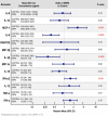
Figure 1
Figure 1
Comparisons of concentrations of 13 cytokines, chemokines and innate immune factors in endocervical fluids from Controls (n=24) and DMPA users (n=15). Fluids were collected by insertion of an ophthalmic sponge (Merocel) into the endocervical canal for 90 s; fluids were extracted and analyzed on a Milliplex platform as described in Methods section. The specific biomarker is indicated in the left column. The next column shows age-adjusted mean concentration (pg ml−1) and 95% confidence intervals of the corresponding biomarker from Controls and DMPA users, ordered by biomarker concentration in Controls from highest (top) to lowest (bottom). The middle column shows the ratio (circle) and 95% confidence intervals (bars) of concentrations from DMPA users compared with Control for each biomarker. The column in the far right gives specific P-values for the ratios with significant values (P-values <0.05) highlighted in red.
Figure 2
Figure 2
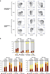
(a) Flow cytometry gating used in the analysis of endocervical and endometrial T-cell phenotypes. After designating a lymphocyte gate based on scatter, followed by doublet discrimination, dead cells were removed by dye exclusion; CD3+CD66b− viable cells (not shown) were then subdivided into CD4+ or CD8+ populations. The resulting T-cell populations were then assessed for expression of three pairs of surface markers, as described in the text. Shown from left to right, these were: differentiation markers CCR7 and CD45RA; activation markers CD38 and HLA-DR; chemokine receptors CCR5 and CXCR4. Quadrant gates were drawn based on fluorescence-minus-one (FMO) controls. Numbers in each quadrant indicate percentages of CD4+ or CD8+ T cells expressing various combinations of markers. Data shown are from a representative participant using DMPA. (b) Flow cytometric phenotyping data. Bivariate distributions of three pairs of phenotypes: (i) CCR7/CD45RA, (ii) CD38/HLADR, and (iii) CXCR4/CCR5, are summarized for CD4+ and CD8+ T cells from endocervical cytobrush and endometrium, as indicated in the headings. The stacked box plots summarize the relative proportions of each phenotype differentiated by color, analyzed as a function of exposure group (control or DMPA) in models stratified by tissue and cell type. P-values are reported for distributional differences between arms as described in Methods section; those values <0.05 are shown in red. Color-coding for each pair of phenotypic markers follows the same pattern in all graphs: +/− red, −/+ yellow, +/+ orange, −/− gray. TEMRA indicates terminally differentiated effector cells expressing CD45RA.
Figure 3
Figure 3
The figure demonstrates differences in macrophage density by immunohistochemical detection in endometrial biopsies from 2 control participants (a and b) and 2 DMPA users (d and e). Five-micron paraffin sections of formalin-fixed endometrial biopsies were incubated with mouse monoclonal anti-CD68 antibodies (a, b, d and e). Background staining was measured by incubation with an immunoglobulin IgG1 isotype control (1:50) matching the anti-CD68 isotype (c and f). Detection of antibody binding was measured by peroxidase-conjugated goat anti-mouse secondary antibodies and diaminobenzidine substrate, as described in Methods section. Slides were counterstained with hematoxylin. Dark brown nuclear staining reflects the presence of macrophages. Bars=50 μm.

References

    1. Ralph, L.J., McCoy, S.I., Shiu, K. & Padian, N.S. Hormonal contraceptive use and women’s risk of HIV acquisition: a meta-analysis of observational studies. Lancet Infect. Dis. 15, 181–189 (2015). - PMC - PubMed
    1. Butler, A.R., Smith, J.A., Polis, C.B., Gregson, S., Stanton, D. & Hallett, T.B. Modelling the global competing risks of a potential interaction between injectable hormonal contraception and HIV risk. Aids 27, 105–113 (2013). - PMC - PubMed
    1. Morrison, C.S. et al. Hormonal contraception and the risk of HIV acquisition: an individual participant data meta-analysis. PLoS Med. 12, e1001778 (2015). - PMC - PubMed
    1. Hormonal Contraceptive Methods for Women at High Risk of HIV and Living with HIV, World Health Organization: Geneva, (2014). - PubMed
    1. Colvin, C.J. & Harrison, A. Broadening the debate over HIV and hormonal contraception. Lancet Infect. Dis. 15, 135–136 (2015). - PMC - PubMed

Publication types