. 2017 Jan 4:8:13932.
doi: 10.1038/ncomms13932.

Evolutionary conservation and in vitro reconstitution of microsporidian iron-sulfur cluster biosynthesis

Affiliations

Evolutionary conservation and in vitro reconstitution of microsporidian iron-sulfur cluster biosynthesis

Sven-A Freibert et al. Nat Commun. .

Abstract

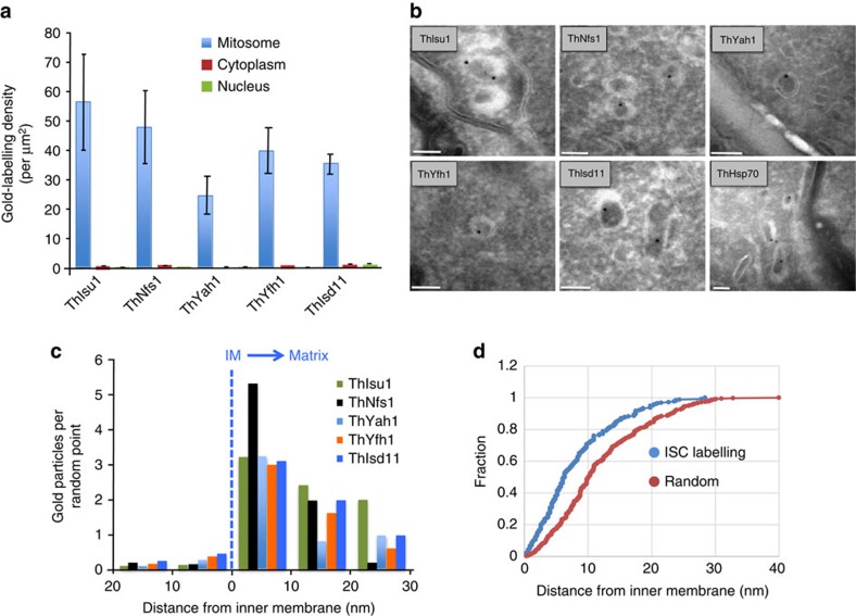
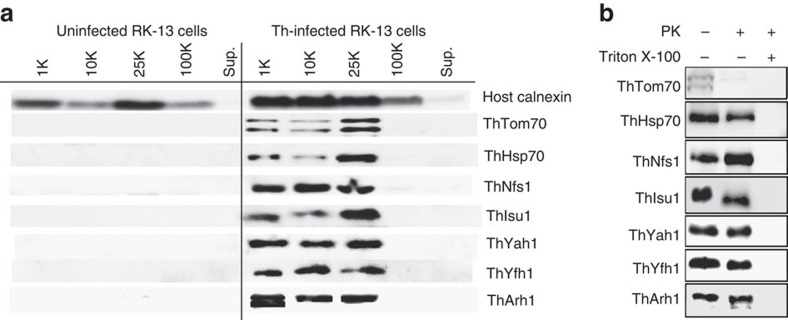
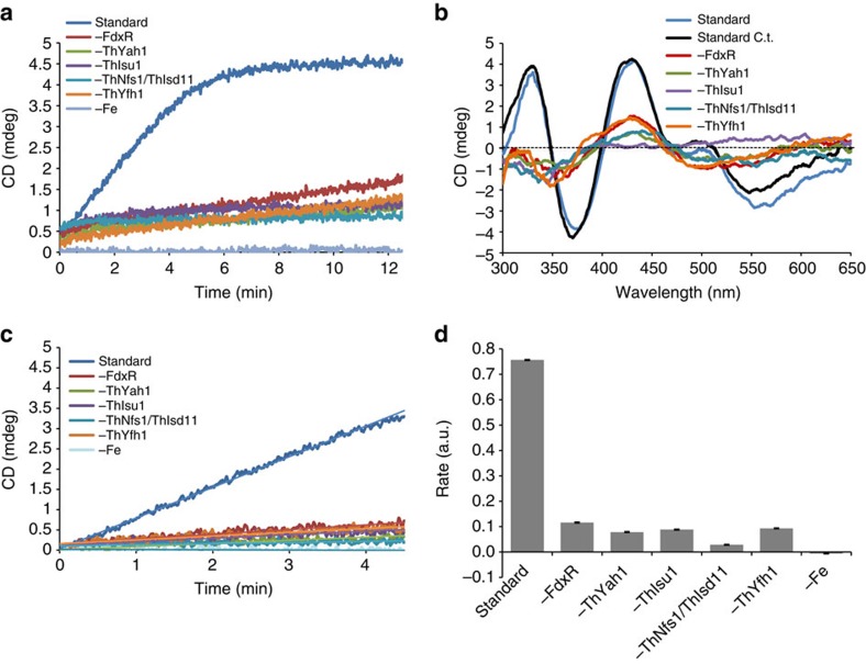
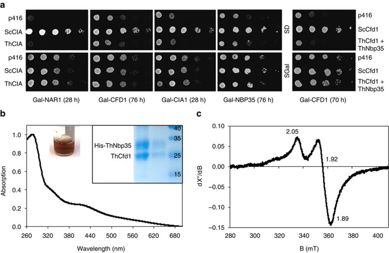
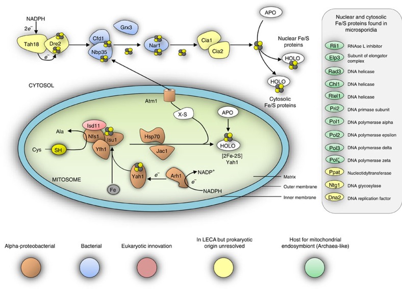
Microsporidians are obligate intracellular parasites that have minimized their genome content and sub-cellular structures by reductive evolution. Here, we demonstrate that cristae-deficient mitochondria (mitosomes) of Trachipleistophora hominis are the functional site of iron-sulfur cluster (ISC) assembly, which we suggest is the essential task of these organelles. Cell fractionation, fluorescence imaging and immunoelectron microscopy demonstrate that mitosomes contain a complete pathway for [2Fe-2S] cluster biosynthesis that we biochemically reconstituted using purified mitosomal ISC proteins. The T. hominis cytosolic iron-sulfur protein assembly (CIA) pathway includes the essential Cfd1-Nbp35 scaffold complex that assembles a [4Fe-4S] cluster as shown by spectroscopic methods in vitro. Phylogenetic analyses reveal that the ISC and CIA pathways are predominantly bacterial, but their cytosolic and nuclear target Fe/S proteins are mainly archaeal. This mixed evolutionary history of Fe/S-related proteins and pathways, and their strong conservation among highly reduced parasites, provides compelling evidence for the ancient chimeric ancestry of eukaryotes.


Figures

Figure 1
Figure 1. Sub-cellular localization of T. hominis Fe/S-cluster assembly components.
Thawed cryo-sections of glutaraldehyde-fixed T. hominis-infected RK-13 cells were labelled with antibodies to T. hominis ISC components and protein-A gold. (a) Quantitative analysis. Labelling is expressed as density of gold labelling over compartment profile area (estimated by point counting; see ‘Methods' section; 34–40 micrographs analysed per protein/experiment with following number of golds per experiment: ThIsu1, 64; ThNfs1, 67; ThYah1, 35; ThYfh1, 70; ThIsd11, 111). Error bars indicate the s.e.m. (N=3). (b) Images illustrating the distribution of labelling for ISC components in mitosomes (these show examples of positive labelling only and therefore do not reflect densities quantified in a). Labelling appears to be located over the matrix of the double membrane-bounded organelle profiles (with mean minor and major axes of 80 and 127 nm, respectively; N=50 mitosome profiles). The analysed profiles were morphologically undistinguishable from mitosomes that labelled positively for ThHsp70 as shown here and described previously. Bars=100 nm. (c) The distribution of immunogold labelling for the indicated ISC proteins over mitosome profiles was compared with an equivalent number of random points. (IM, inner mitosomal membrane location). Labelling was expressed as gold particles per random point count and indicates the concentration of labelling at the IM/matrix interface. The numbers of gold and random counts were as follows: ThIsu1, 79; ThNfs1, 66; ThYah1, 37; ThYfh1, 63; ThIsd11, 50. Labelling for all tested ISC components was towards the mitosomal matrix with an enrichment at the IM. (d) Immunogold labelling distribution over mitosome matrix. Cumulative fraction plot shows pooled gold labelling data for all five ISC components (ISC labelling) compared with points located simple uniform random (random). ISC components show relative enrichment of labelling close to the IM (see Supplementary Fig. 3 for individual analyses).
Figure 2
Figure 2. Biochemical localization of the T. hominis ISC pathway components.
(a) Western blots using antibodies to T. hominis proteins for fractions obtained by differential centrifugation from RK cells or RK cells infected with T. hominis (Th). Centrifugation speeds of pellet fractions are given above each lane. Sup=final 100,000g (100 K) supernatant. (b) Western blot of the 25,000g (25 K) pellet fraction of T. hominis-infected RK cells treated with or without proteinase K (PK) and Triton X-100 detergent as indicated.
Figure 3
Figure 3. In vitro reconstitution of Fe/S-cluster synthesis on T. hominis Isu1.
(a) The T. hominis proteins ThIsu1, ThNfs–ThIsd11, ThYfh1, ThYah1 and human FdxR (the homologue of yeast Arh1) were mixed anaerobically in buffer R (standard reaction). Cysteine was added to start Fe/S-cluster synthesis, which was recorded by the CD signal change at 431 nm. Replicate reactions were performed, in which individual components of the T. hominis core ISC pathway or iron (Fe) were systematically omitted. (b) After 15 min full CD spectra were recorded for each reaction mixture, and compared with the spectrum of a standard reaction using ISC proteins from C. thermophilum (C.t.). (c) The initial rates of Fe/S-cluster synthesis on ThIsu1 for the different reactions were recorded. (d) The initial rates were estimated by linear regression and compared with the standard reaction using C.t. ISC components. N≥5; Error bars=s.d.
Figure 4
Figure 4. Localization of the T. hominis homologue of mitochondrial Atm1.
(a) Fractionation by differential ultracentrifugation was carried out on healthy and T. hominis-infected RK cells and the fractions were immunostained using an antibody to ThAtm1_1. (b) Proteinase K (PK) protection was performed on the mitosome-enriched 25,000g (25 K) fraction of infected cells, with Triton X-100 (TX) used to solubilize the membranes. A Coomassie-stained gel of the fractions after the protection assay is shown on the right. (ci) Immunofluorescence of fixed RK-13 cells infected with T. hominis using antibodies raised against ThHsp70 (c,e; green; rat) or ThAtm1_1 (d,e; red; rabbit). DAPI was used to label host and parasite nuclear DNA (blue). The ThAtm1_1 antiserum was pre-purified using uninfected RK-13 cell lysate immobilized on nitrocellulose following SDS–PAGE. Colocalized pixels were identified using Zeiss Axiovision software, and are pseudo-coloured pink as shown in f. The DIC image in (g) shows the individual meronts in the infected RK cell. (h,i) A close-up of a different sample of meronts showing the typical distribution of the signals for the two antibodies. (j) Colocalisation scatter plots against the different channels were generated by Axiovision software and the antibody to the protein import receptor ThTom70 was used as a positive control for colocalisation with ThHsp70. The scatter plots for ThAtm1_1 with ThHsp70 and ThTom70 with ThHsp70 are similar, indicating co-localization of both proteins with ThHsp70. (km), The colocalized pixel counts were also quantified (n=3, error bars=s.d.) and represented graphically for ThHsp70 and either (k) ThAtm1_1 or (l) ThTom70. (m) The extent of mitosomal labelling by the ThAtm1_1 antibody relative to other cell compartments was quantified based on the proportion of pixel intensity across six fields of view for each of three replicates (error bars=s.d.).
Figure 5
Figure 5. Functional reconstitution of the microsporidian CIA scaffold complex.
(a) Yeast complementation assay. Functionality of the T. hominis CIA components ThNar1, ThCfd1, ThCia1 or ThNbp35 was tested by their ability to rescue the growth defect of respective regulatable Gal-CIA yeast mutants on glucose-containing minimal medium (s.d.). Indicated cells were transformed with plasmid p416-MET25 containing either no gene (p416), the respective yeast (Sc) or the homologous T. hominis (Th) CIA genes. Gal-CFD1 cells were additionally transformed with p415-MET25 to allow co-production of both ThCfd1 and ThNbp35 (right). Cells were depleted of the respective nuclear-encoded CIA proteins by growth on s.d. medium for the indicated times at 30 °C. Serial tenfold dilutions were spotted onto agar plates containing either SGal (minimal medium plus galactose) or s.d. medium, and growth was continued for 2 days at 30 °C. None of the T. hominis genes improved growth of the yeast CIA protein-depleted cells. (b) T. hominis ThCfd1 and His-tagged ThNbp35 were co-expressed in E. coli and co-purified as a complex by affinity chromatography (insight, right). Fe/S clusters were chemically reconstituted on the ThCfd1–HisThNbp35 complex resulting in a dark-brown protein solution (inset, left). Ultraviolet–vis spectroscopy revealed an absorption peak around 420 nm indicative of the formation of [4Fe–4S] clusters on the complex. (c) The presence of [4Fe–4S] clusters was confirmed by X-band EPR spectroscopy of a reduced sample (40 μM, treated with 0.2 mM Na-dithionite) recorded at 10 K. Experimental conditions: frequency 9.6359 GHz, power 1 mW, modulation 0.75 mT/100 kHz. The numbers are the principal g values obtained by simulation.
Figure 6
Figure 6. Evolutionary origins of microsporidian Fe/S-related proteins.
The cartoon shows the components of the eukaryotic ISC and CIA machineries conserved on the genomes of E. cuniculi and T. hominis. Individual components are coloured according to their inferred evolutionary origins (Supplementary Fig. 2). Components of the mitosomal ISC pathway appear to have originated from the mitochondrial endosymbiont. The CIA pathway is largely bacterial in character, and not archaeal as might be expected based on evidence for an archaeal origin of the host for the mitochondrial endosymbiosis. By contrast, important nuclear and cytosolic Fe/S proteins do appear to have an archaeal origin. Monophyly of eukaryotic sequences, including those from microsporidians, is generally observed, suggesting that there is strong negative selection against gene replacement due to the important roles that Fe/S proteins play in eukaryotic physiology.

References

    1. Vavra J. & Lukes J. Microsporidia and ‘the art of living together'. Adv. Parasitol. 82, 253–319 (2013). - PubMed
    1. Nakjang S. et al.. Reduction and expansion in microsporidian genome evolution: new insights from comparative genomics. Genome Biol. Evol. 5, 2285–2303 (2013). - PMC - PubMed
    1. Williams B. A., Hirt R. P., Lucocq J. M. & Embley T. M. A mitochondrial remnant in the microsporidian Trachipleistophora hominis. Nature 418, 865–869 (2002). - PubMed
    1. Katinka M. D. et al.. Genome sequence and gene compaction of the eukaryote parasite Encephalitozoon cuniculi. Nature 414, 450–453 (2001). - PubMed
    1. Embley T. M. & Martin W. Eukaryotic evolution, changes and challenges. Nature 440, 623–630 (2006). - PubMed

Publication types

MeSH terms