. 2017 Jan 4:7:39998.
doi: 10.1038/srep39998.

Mechanisms of Yersinia YopO kinase substrate specificity

Affiliations

Mechanisms of Yersinia YopO kinase substrate specificity

Wei Lin Lee et al. Sci Rep. .

Abstract

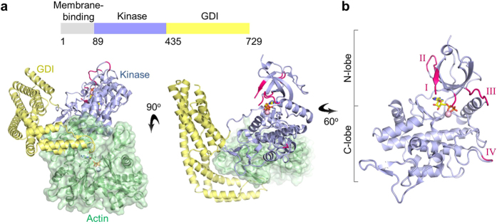
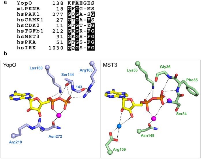
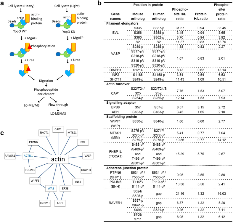
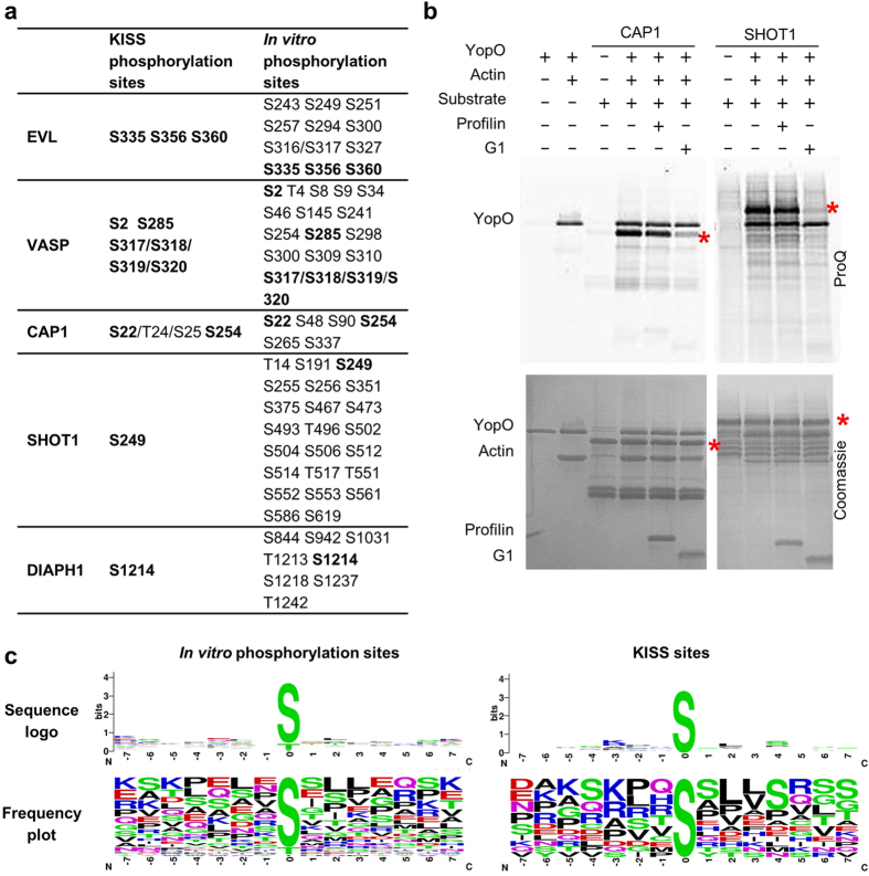
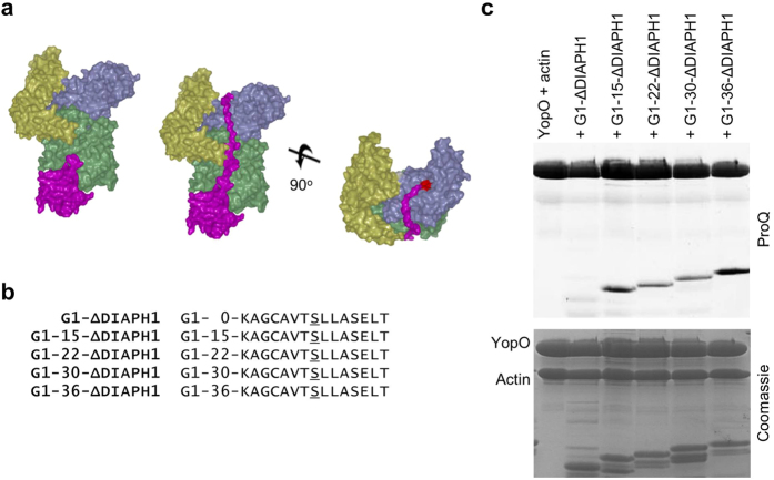
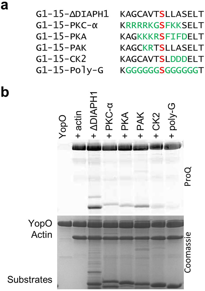
Yersinia bacteria cause a range of human diseases, including yersiniosis, Far East scarlet-like fever and the plague. Yersiniae modulate and evade host immune defences through injection of Yersinia outer proteins (Yops) into phagocytic cells. One of the Yops, YopO (also known as YpkA) obstructs phagocytosis through disrupting actin filament regulation processes - inhibiting polymerization-promoting signaling through sequestration of Rac/Rho family GTPases and by using monomeric actin as bait to recruit and phosphorylate host actin-regulating proteins. Here we set out to identify mechanisms of specificity in protein phosphorylation by YopO that would clarify its effects on cytoskeleton disruption. We report the MgADP structure of Yersinia enterocolitica YopO in complex with actin, which reveals its active site architecture. Using a proteome-wide kinase-interacting substrate screening (KISS) method, we identified that YopO phosphorylates a wide range of actin-modulating proteins and located their phosphorylation sites by mass spectrometry. Using artificial substrates we clarified YopO's substrate length requirements and its phosphorylation consensus sequence. These findings provide fresh insight into the mechanism of the YopO kinase and demonstrate that YopO executes a specific strategy targeting actin-modulating proteins, across multiple functionalities, to compete for control of their native phospho-signaling, thus hampering the cytoskeletal processes required for macrophage phagocytosis.


Figures

Figure 1
Figure 1. Crystal structure of YopO-actin:MgADP.
(a) Crystal structure of the kinase (light blue) and GDI (pale yellow) domains in complex with actin (pale green). MgADP lies in the active site of the YopO kinase domain. (b) Structure of the kinase domain of YopO. Regions that are ordered in this ADP-bound structure, but were disordered in the apo structure (PDB: 4CI6), are colored pink and labeled I–IV with reference to Supplementary Fig. 1.
Figure 2
Figure 2. Coordination of ADP by the non-consensus YopO P-loop.
(a) Sequence alignment of P-loop sequences across kinases from different families. Full alignments can be found in Supplementary Fig. 2. (b) Hydrogen-bonding interactions that coordinate the kinase-bound ADP. Magenta spheres represent Mg2+ ion in YopO and Mn2+ ions in MST3 (PDB: 3A7J) while blue spheres represent water. For clarity, only the side chain is shown for some residues while their attachment to the main chain is represented as a sphere, and the side-chain atoms of residue 143 are not shown.
Figure 3
Figure 3. Screening for YopO substrates using the KISS method.
(a) Scheme of the KISS method. (b) Proteins identified as YopO kinase substrates. Phosphorylation sites with phosphosite/peptide ratio above 2 were considered as putative YopO phosphorylation sites. The phosphosites were further subjected to cut-offs of mass spectrometry identification score of 44 and protein ratio count of 4. Alternative names for the proteins were shown in brackets. KISS was performed using mouse monocyte/macrophage cell line Raw264.7. Phosphorylation sites and their corresponding residues in the human ortholog are shown. p denotes reported phosphorylation of the human or mouse ortholog according to Phosphosite. # denotes ambiguous identification in MS due to close proximity of potential phosphorylation sites. (c) Model of the network of direct and indirect interactions of YopO substrates to actin. Proteins shown in blue were detected in KISS but were not found to be significantly phosphorylated.
Figure 4
Figure 4. In vitro phosphorylation of actin-binding proteins by YopO.
(a) Mass spectrometry identification of phosphorylation sites obtained by in vitro phosphorylation. Human EVL, VASP and SHOT1 and mouse CAP1 and DIAPH1 (residues 583–1255) were used. Residues numbering follows the mouse sequences for clarity. (b) In vitro phosphorylation assay, monitored by sequential ProQ Diamond phosphoprotein and Coomassie staining. Red asterisks indicate the molecular weights of the substrates. Final protein concentrations used in the assay were YopO (1.1 μM), G-actin (1.1 μM), substrates (5.5 μM), G1 (3.3 μM) and profilin (3.3 μM). (c) Preferred YopO phosphorylation motifs, computed from sequences in (Supplementary Fig. 9).
Figure 5
Figure 5. Length requirement from actin-binding site to kinase catalytic cleft.
(a) Model for phosphorylation of recruited actin-binding proteins by YopO. Model 1 is a superimposition of gelsolin domain 1 up to FKHV (28–152) from the G1-G3:actin complex (PDB:3FFK). Model in middle and right show an extension of 26 residues reaching up into the catalytic cleft of YopO with active site residues in red. (b) Artificial substrates of YopO with G1 fused to a phosphorylation sequence from DIAPH1 with the insertion of poly glycine-alanine linkers of different lengths between G1 and the phosphorylation sequence. (c) In vitro phosphorylation assay of artificial substrates of various lengths. The SDS-PAGE gel was sequentially imaged with ProQ Diamond phosphoprotein and Coomassie staining. Final protein concentrations were YopO (1.8 μM), G-actin (1.8 μM), artificial substrates (5.4 μM).
Figure 6
Figure 6. Recognition of phosphorylation motifs of other kinases by YopO.
(a) Artificial substrates containing phosphorylation motifs belonging to different kinases. (b) In vitro phosphorylation assay. The SDS-PAGE was sequentially imaged with ProQ Diamond phosphoprotein and Coomassie staining. Final protein concentrations were YopO (1.8 μM), G-actin (1.8 μM), artificial substrates (5.4 μM).

References

    1. Cornelis G. R. et al.. The Virulence Plasmid of Yersinia, an Antihost Genome. Microbiol Mol Biol Rev 62, 1315–1352 (1998). - PMC - PubMed
    1. Viboud G. I. & Bliska J. B. Yersinia outer proteins: Role in Modulation of Host Cell Signaling Responses and Pathogenesis. Annu Rev Microb 59, 69–89 (2005). - PubMed
    1. Hamid N. et al.. YopH dephosphorylates Cas and Fyn-binding protein in macrophages. Microb Pathog 27, 231–242 (1999). - PubMed
    1. Shao F. et al.. Biochemical characterization of the Yersinia YopT protease: Cleavage site and recognition elements in Rho GTPases. Proc Natl Acad Sci 100, 904–909 (2003). - PMC - PubMed
    1. Black D. S. & Bliska J. B. The RhoGAP activity of the Yersinia pseudotuberculosis cytotoxin YopE is required for antiphagocytic function and virulence. Mol Microbiol 37, 515–527 (2000). - PubMed

Publication types

MeSH terms

LinkOut - more resources