

Continuous fabrication of nanostructure arrays for flexible surface enhanced Raman scattering substrate
- PMID: 28051175
- PMCID: PMC5209699
- DOI: 10.1038/srep39814
Continuous fabrication of nanostructure arrays for flexible surface enhanced Raman scattering substrate
Abstract
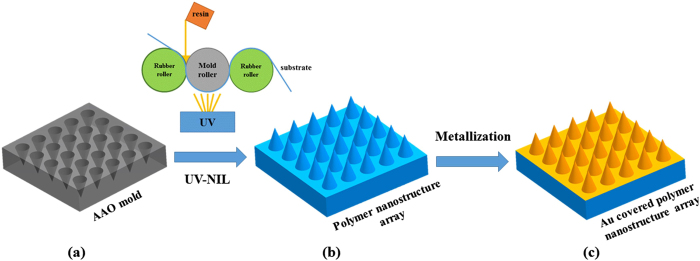
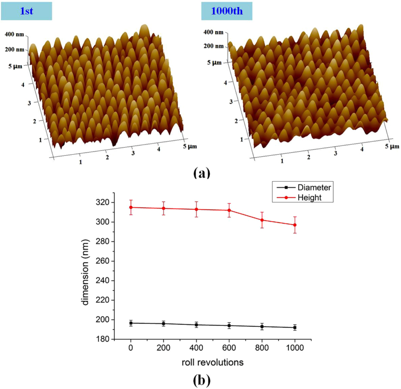
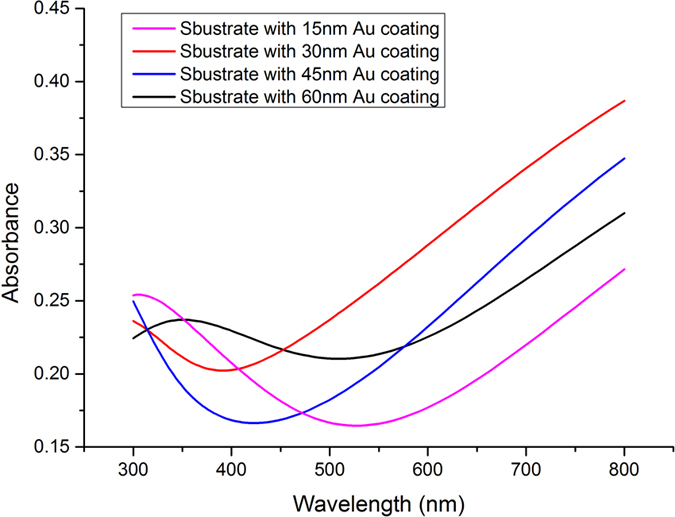
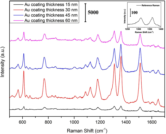
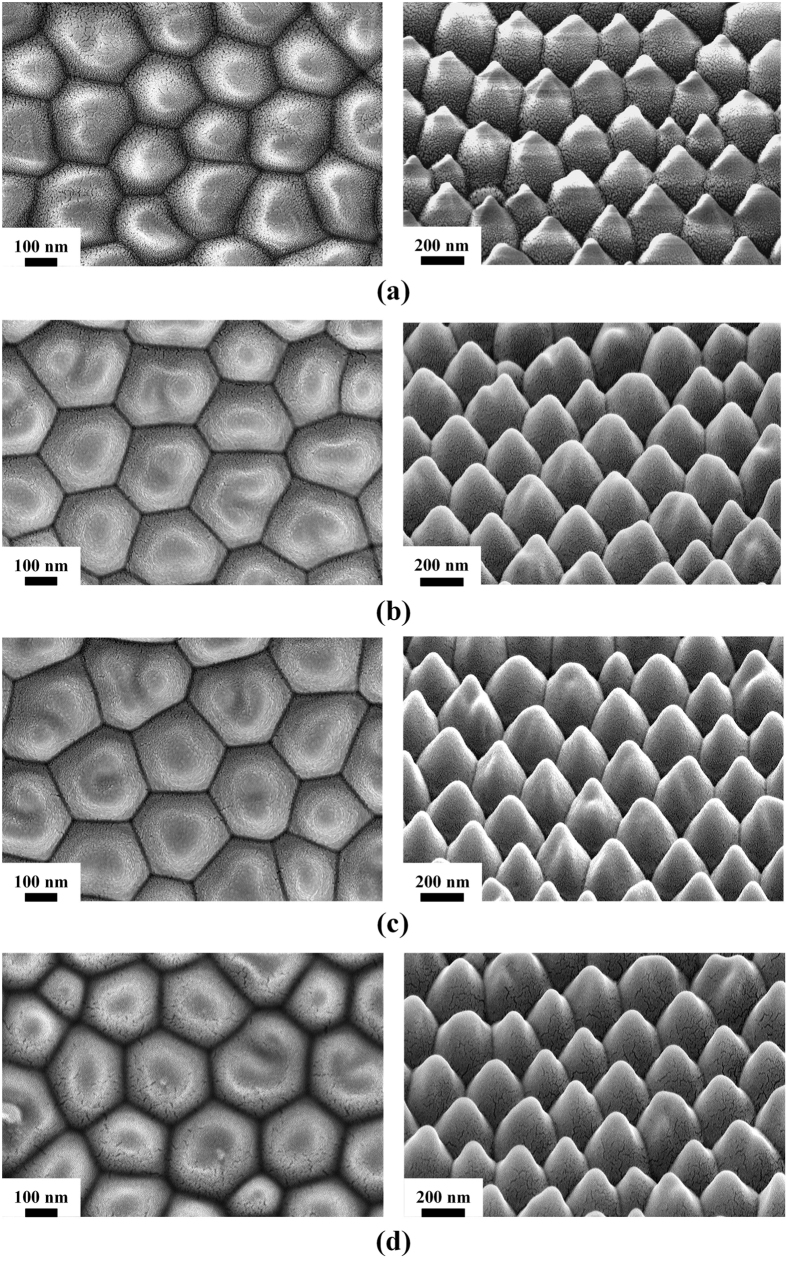
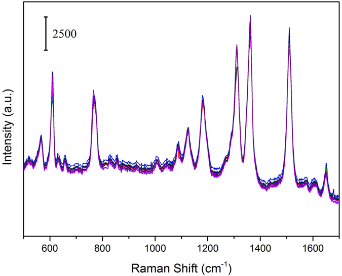
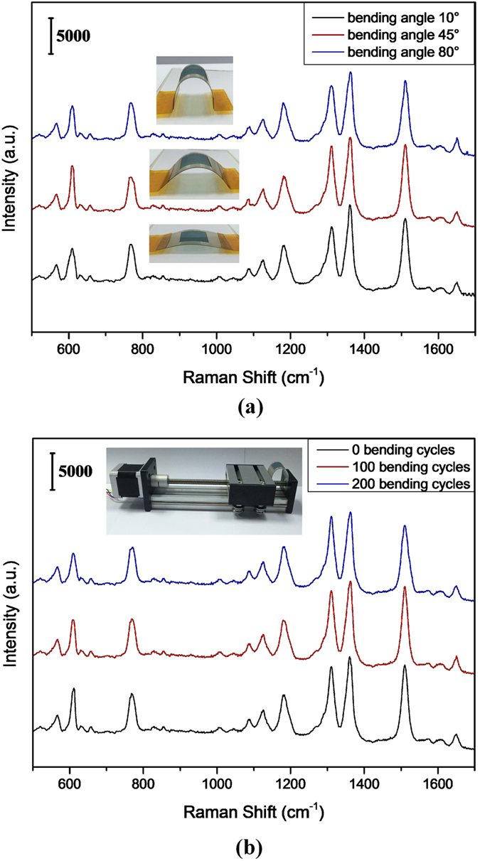
Surface-enhanced Raman spectroscopy (SERS) has been a powerful tool for applications including single molecule detection, analytical chemistry, electrochemistry, medical diagnostics and bio-sensing. Especially, flexible SERS substrates are highly desirable for daily-life applications, such as real-time and in situ Raman detection of chemical and biological targets, which can be used onto irregular surfaces. However, it is still a major challenge to fabricate the flexible SERS substrate on large-area substrates using a facile and cost-effective technique. The roll-to-roll ultraviolet nanoimprint lithography (R2R UV-NIL) technique provides a solution for the continuous fabrication of flexible SERS substrate due to its high-speed, large-area, high-resolution and high-throughput. In this paper, we presented a facile and cost-effective method to fabricate flexible SERS substrate including the fabrication of polymer nanostructure arrays and the metallization of the polymer nanostructure arrays. The polymer nanostructure arrays were obtained by using R2R UV-NIL technique and anodic aluminum oxide (AAO) mold. The functional SERS substrates were then obtained with Au sputtering on the surface of the polymer nanostructure arrays. The obtained SERS substrates exhibit excellent SERS and flexibility performance. This research can provide a beneficial direction for the continuous production of the flexible SERS substrates.
Figures
References
-
- Fleischmann M., Hendra P. J. & McQuillan A. Raman spectra of pyridine adsorbed at a silver electrode. Chem. Phys. Lett. 26, 163–166 (1974).
-
- McNay G., Eustace D., Smith W. E., Faulds K. & Graham D. Surface-enhanced Raman scattering (SERS) and surface-enhanced resonance Raman scattering (SERRS): a review of applications. Appl. Spectrosc. 65, 825–837 (2011). - PubMed
-
- Sharma B., Frontiera R. R., Henry A. I., Ringe E. & Van Duyne R. P. SERS: materials, applications, and the future. Materials today 15, 16–25 (2012).
-
- Chen J. et al. Multiple myeloma detection based on blood plasma surface-enhanced Raman spectroscopy using a portable Raman spectrometer. Laser Phys. Lett. 13, 105601 (2016).
-
- Zou Y. et al. Urine surface-enhanced Raman spectroscopy for non-invasive diabetic detection based on a portable Raman spectrometer. Laser Phys. Lett. 13, 065604 (2016).
Publication types
LinkOut - more resources
Full Text Sources
Other Literature Sources
Miscellaneous
