. 2017 Jan 4:7:40067.
doi: 10.1038/srep40067.

The larvicide pyriproxyfen blamed during the Zika virus outbreak does not cause microcephaly in zebrafish embryos

Affiliations

The larvicide pyriproxyfen blamed during the Zika virus outbreak does not cause microcephaly in zebrafish embryos

Stefania Dzieciolowska et al. Sci Rep. .

Abstract

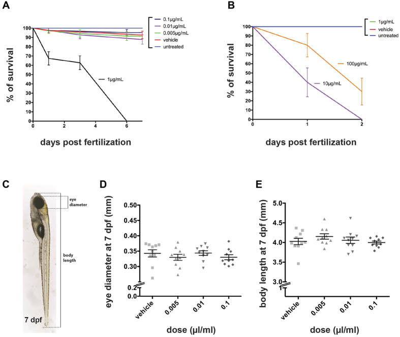
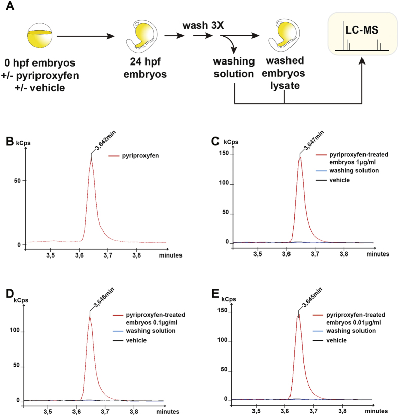
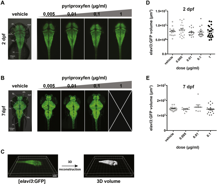
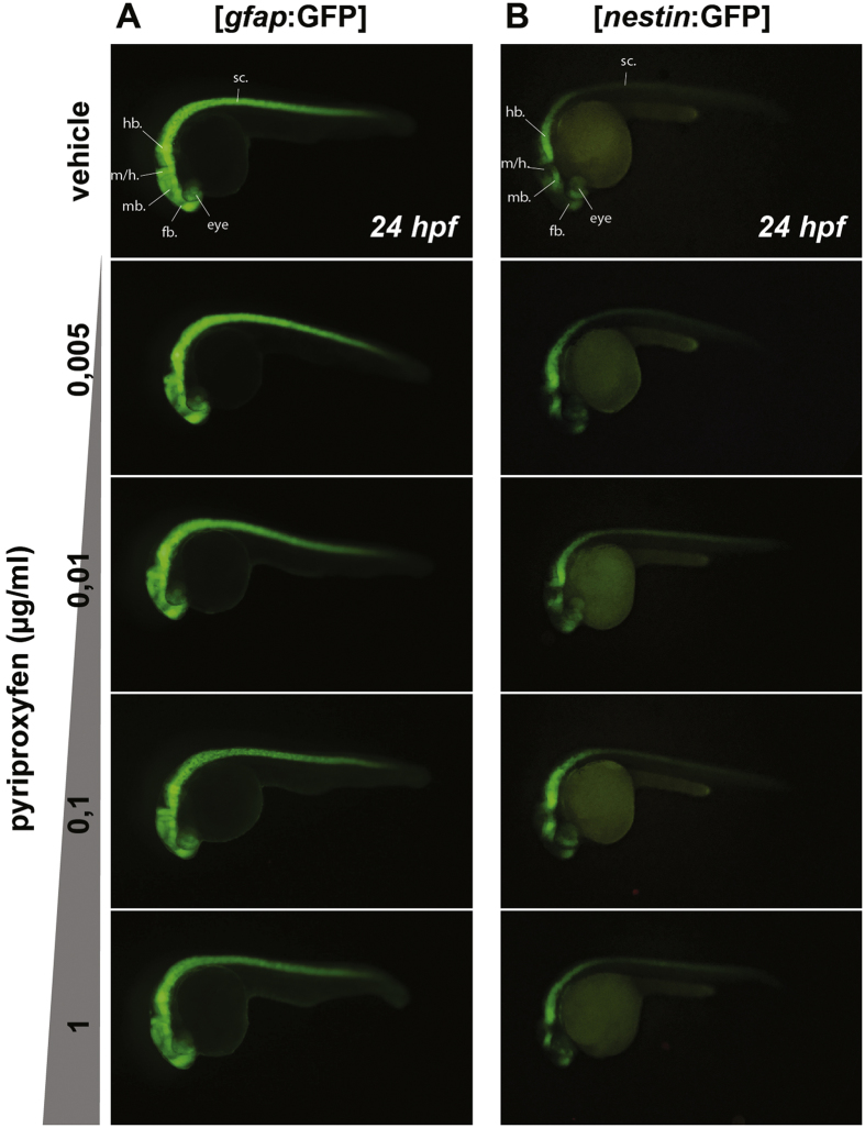
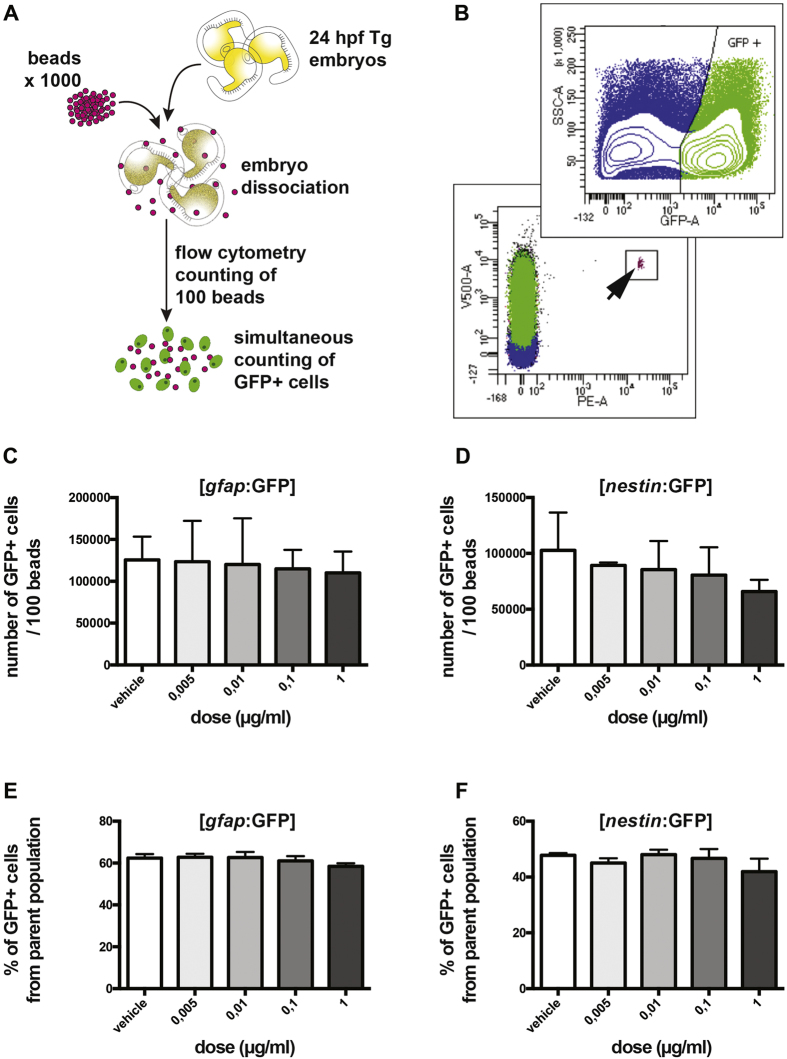
Although the zika virus (ZIKV) has now been strongly correlated with emerging cases of microcephaly in the Americas, suspicions have been raised regarding the use of pyriproxyfen, a larvicide that prevents mosquito development, in drinking water. The effects of this compound on neurodevelopment have not yet been addressed specifically in vertebrates. As a result, we aimed at addressing the effects, if any, of pyriproxyfen on neurodevelopment in the zebrafish embryo as a vertebrate model. Using zebrafish transgenic lines expressing GFP in different cell populations (elavl3 in newborn neurons, gfap and nestin in neural stem cells), we focused on the analysis of whole embryonic brain volume after confocal 3D-reconstruction and the quantification of purified neural stem cells during early neurodevelopment by FACS-cell sorting from whole in vivo embryos. Interestingly, though lethal at very high doses, pyriproxyfen did not cause brain malformation nor any significant changes in the number of observed stem cells in the developing central nervous system. Our data indicate that pyriproxyfen does not affect central nervous system development in zebrafish, suggesting that this larvicide on its own, may not be correlated with the increase in microcephaly cases reported recently.


Figures

Figure 1
Figure 1. Pyriproxyfen is toxic at high doses.
(A) Percentage of survival of embryos treated from 2–4 cell stage with various pyriproxyfen concentrations ranging from 0,005 μg/ml to 1 μg/ml. The latest dose appears to be lethal with 100% of death after 5 days of development. Lower doses of pyriproxyfen (0,005; 0,01; 0,1 μg/ml) do not induce a severe decrease in the survival rate (N = 2 batches, n = 20 embryos per condition). (B) Higher doses of pyriproxyfen (10 and 100 μg/ml) are lethal as early as 2 dpf (N = 2, n = 20). (C) Representation of measurements performed on 7 dpf larvae to assess gross morphology effects. (D,E) Eye diameter and body length at 7 dpf of pyriproxyfen-treated embryos are not significantly different from vehicle (EtOH)- treated embryos (p > 0.05) (n = 10 per condition).
Figure 2
Figure 2. Liquid chromatography-mass spectrometry detection of pyriproxyfen in treated embryos.
(A) Steps involved in the preparation for pyriproxyfen extraction of embryos (hpf: hours post-fertilization). (B) LC chromatogram of commercial pyriproxyfen at a concentration of 1.56 nM eluting at a retention time of 3.64 min. (CE) LC chromatogram illustrating pyriproxyfen extraction from embryos treated with different concentrations of the molecule compared to vehicle treated embryos and the 3rd washing solution. (Cps: counts per second).
Figure 3
Figure 3. Embryonic and larval brain volumes are not affected by pyriproxyfen treatment.
(A) Transgenic [elavl3:GFP] 2 dpf embryo treated with vehicle, 0,005 μg/ml, 0,01 μg/ml, 0,1 μg/ml and 1 μg/ml pyriproxyfen were imaged under a confocal microscope. The morphology of the embryonic brain is not affected by any of the doses. (B) Transgenic [elavl3:GFP] 7 dpf larvae treated with vehicle, 0,005 μg/ml, 0,01 μg/ml, 0,1 μg/ml pyriproxyfen were imaged under a confocal microscope. Of note is that none of the embryos treated with a dose of 1 μg/ml survived past 6 dpf. The morphology of the larval brain is not affected by any of the doses. (C) 3D-volume reconstruction of [elavl3:GFP] embryos using Imaris software (Bitplane) and confocal microscopy. (D,E) Quantification of embryonic (2 dpf) (D) and larval (7 dpf) (E) brain volumes shows no significant differences between vehicle-treated and pyriproxyfen-treated embryos (p > 0.05). (N = 2, n = 7 per condition). sc.: spinal cord; hb.: hindbrain; m/h: midbrain/hindbrain boundary; mb.: midbrain; fb.: forebrain; ot.: optic tectum; tv.: tectum ventricle; ob.: olfactory bulb.
Figure 4
Figure 4. Neural stem cells regional expression pattern is not affected by pyriproxyfen exposure.
24 hpf transgenic embryos expressing the GFP under the gfap promoter [gfap:GFP] (A) or nestin promoter [nestin:GFP] (B) were treated with different doses of pyriproxyfen from the 2–4 cell stage onwards. Whole GFP expression pattern of both transgenic lines is not affected by the treatment. (N = 2, n = 25 per condition). sc.: spinal cord; hb.: hindbrain; m/h: midbrain/hindbrain boundary; mb.: midbrain; fb.: forebrain.
Figure 5
Figure 5. Neural stem cell populations are not affected by pyriproxyfen exposure.
(A) Whole 24 hpf transgenic embryos ([gfap:GFP] or [nestin:GFP] were dissociated into single-cells in a medium containing a 1,000 fluorescent beads. 100 beads were counted by flow cytometry and the number of GFP+ cells was counted simultaneously. (B) The GFP+ cell population ([gfap:GFP] embryos) was distinguishable using a 488 nm laser with a 530/30 BP filter (upper panel). The fluorescent beads were distinguishable using a 561 nm laser with a 610/20 BP filter and a 405 nm laser with a 525/50 BP filter. The beads are completely separated from GFP+ cells on a FSC/SSC dot plot (lower panel, arrow). (C,D) The number of gfap+ cells and nestin+ counted per 100 beads was not significantly different upon pyriproxyfen exposure when compared to vehicle-treated embryos (p > 0.05). (E,F) The same results presented as a percentage of GFP+ cells/parent population also show no significant differences upon pyriproxyfen exposure among both gfap+ cells and nestin+ cells (p > 0.05). (N = 2, n = 25 per condition).

Similar articles

Cited by

References

    1. Lazear H. M., Stringer E. M. & de Silva A. M. The Emerging Zika Virus Epidemic in the Americas: Research Priorities. JAMA: the journal of the American Medical Association 315, 1945–1946, doi: 10.1001/jama.2016.2899 (2016). - DOI - PubMed
    1. Gatherer D. & Kohl A. Zika virus: a previously slow pandemic spreads rapidly through the Americas. J Gen Virol 97, 269–273, doi: 10.1099/jgv.0.000381 (2016). - DOI - PubMed
    1. Duffy M. R. et al.. Zika virus outbreak on Yap Island, Federated States of Micronesia. The New England journal of medicine 360, 2536–2543, doi: 10.1056/NEJMoa0805715 (2009). - DOI - PubMed
    1. Oliveira Melo A. S. et al.. Zika virus intrauterine infection causes fetal brain abnormality and microcephaly: tip of the iceberg? Ultrasound Obstet Gynecol 47, 6–7, doi: 10.1002/uog.15831 (2016). - DOI - PubMed
    1. Corona-Rivera J. R. et al.. Report and review of the fetal brain disruption sequence. Eur J Pediatr 160, 664–667 (2001). - PubMed

Publication types

Grants and funding

LinkOut - more resources