. 2017 Jan 4:8:14034.
doi: 10.1038/ncomms14034.

Architecture and mechanism of the late endosomal Rab7-like Ypt7 guanine nucleotide exchange factor complex Mon1-Ccz1

Affiliations

Architecture and mechanism of the late endosomal Rab7-like Ypt7 guanine nucleotide exchange factor complex Mon1-Ccz1

Stephan Kiontke et al. Nat Commun. .

Abstract

The Mon1-Ccz1 complex (MC1) is the guanine nucleotide exchange factor (GEF) for the Rab GTPase Ypt7/Rab7 and is required for endosomal maturation and fusion at the vacuole/lysosome. Here we present the overall architecture of MC1 from Chaetomium thermophilum, and in combining biochemical studies and mutational analysis in yeast, we identify the domains required for catalytic activity, complex assembly and localization of MC1. The crystal structure of a catalytic MC1 core complex bound to Ypt7 provides mechanistic insight into its function. We pinpoint the determinants that allow for a discrimination of the Rab7-like Ypt7 over the Rab5-like Vps21, which are both located on the same membrane. MC1 shares structural similarities with the TRAPP complex, but employs a novel mechanism to promote nucleotide exchange that utilizes a conserved lysine residue of Ypt7, which is inserted upon MC1 binding into the nucleotide-binding pocket of Ypt7 and contributes to specificity.


Figures
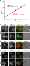
Figure 1
Figure 1. Catalytic activity and localization requirements of Mon1–Ccz1.
(a) Nucleotide exchange rates of Ypt7 are plotted as a function of CtMC1 concentrations. The catalytic efficiency of full-length CtMC1 and a truncated CtMC1core complex (Mon1 195–355, Ccz1 1–249) are comparable. Error bars represent s.d. of three independent biological repeats. (b) GFP-tagged truncations of ScMon1 and (c) ScCcz1 are introduced into mon1Δ and ccz1Δ yeast knockout strains. The N-terminus of Mon1 is dispensable, but deletion of the Mon1 and Ccz1 C-termini cause mislocalization and vacuolar fragmentation. (d) The C-terminus of Mon1 is replaced by a PI3P-binding FYVE domain and artificially recruited to endosomal and vacuolar membranes as seen in a wild-type background, but Mon1-ΔC-FVYE is not able to complement a mon1Δ strain. Scale bars: 5 μm.
Figure 2
Figure 2. Crystal structure of the catalytic CtMC1core complex bound to nucleotide-free CtYpt7–N125I.
(a) The content of the asymmetric unit is shown, comprising two copies of Ypt7 and two Mon1–Ccz1 heterodimers that interact via the domain-swapped helix α3 of Mon1. (b) Stereo image of the 2FO–FC electron density map at 1.8σ contour level of the switch I, switch II and P-loop region of CtYpt7–N125I bound to MC1. (c) Complex structures of MC1 with Ypt7 where the domains' swap was corrected to represent the likely functional unit. (d) Complex structure of TRAPP with Ypt1. The different interaction interfaces between GTPase and GEF are marked.
Figure 3
Figure 3. Selectivity of Mon1–Ccz1 for Ypt7 over Vps21.
(a) For CtVps21, CtYpt7 and different mutations, nucleotide exchange rates are plotted as a function of CtMC1core concentration. Error bars represent s.d. of three independent biological repeats. (b) MC1-interacting interface of Ypt7 is shown in blue. Positions that are conserved in the Ypt7 family, but not the Vps21 family, are highlighted. Labels show the mutations from Ypt7- to Vps21-specific residues. Colours correspond to the graphs in b.
Figure 4
Figure 4. Interaction of Mon1–Ccz1 with Ypt7.
(a) Surface representation of the CtMC1 longin heterodimer. Mutations introduced in the interaction interface with Ypt7 are labelled. (b) Nucleotide exchange rates of Ypt7 are plotted as a function of the concentration of CtMC1core wild-type and different mutations. Error bars represent s.d. of three independent biological repeats. Colours of the graphs correspond to highlighted mutations in a. Functionality test of mutations in (c) mon1Δ and (d) ccz1Δ yeast knockout strains. The vacuolar fragmentation phenotype is rescued by the introduction of ScMon1 and ScCcz1, respectively, but not the mutations corresponding to the GEF-deficient mutants described above. Scale bars: 5 μm.
Figure 5
Figure 5. Mechanism of nucleotide exchange by Mon1–Ccz1.
(a) Superposition of ScYpt7 bound to the GTP analogue GNP in magenta with MC1-bound CtYpt7 in blue. The switch I and switch II regions, which undergo major rearrangements, are labelled. (b) Interaction of F33 and Y37 from switch I of Ypt7 with two hydrophobic pockets on MC1. The surface of MC1 is coloured according to hydrophobicity from green (hydrophilic) to grey (hydrophobic). (c) Nucleotide exchange rates of CtYpt7 wild type and the mutants F33A and K38A are plotted as a function of the concentration of CtMC1core. Error bars represent s.d. of three independent biological repeats. (d) Close-up of the nucleotide pocket from ScYpt7-GNP and MC1-bound CtYpt7. The position of switch II D63/64 and P-loop K21 are unaltered. K38 from switch I is inserted into the pocket and clashes with the Mg2+ involved in coordinating nucleotides.
Figure 6
Figure 6. Model of the nucleotide exchange mechanism by Mon1–Ccz1.
(a) F33 of Ypt7 interacts with the guanosine base to stabilize nucleotide binding. Switch I closes the binding pocket with Y37 and K38 solvent exposed. (b) F33 and Y37 are fixed in hydrophobic pockets on Mon1–Ccz1, leading to switch I rearrangement and opening of the nucleotide-binding pocket. In addition, K38 of Ypt7 is inserted into the nucleotide pocket, repelling with its positively charged terminal amine group the Mg2+ cofactor ion from the binding site.

Similar articles

Cited by

References

    1. Jahn R. & Scheller R. H. SNAREs-engines for membrane fusion. Nat. Rev. Mol. Cell Biol. 7, 631–643 (2006). - PubMed
    1. Stenmark H. Rab GTPases as coordinators of vesicle traffic. Nat. Rev. Mol. Cell Biol. 10, 513–525 (2009). - PubMed
    1. Pan X., Eathiraj S., Munson M. & Lambright D. G. TBC-domain GAPs for Rab GTPases accelerate GTP hydrolysis by a dual-finger mechanism. Nature 442, 303–306 (2006). - PubMed
    1. Langemeyer L. et al.. Diversity and plasticity in Rab GTPase nucleotide release mechanism has consequences for Rab activation and inactivation. Elife 3, e01623 (2014). - PMC - PubMed
    1. Ishida M., Oguchi M. E. & Fukuda M. Multiple types of guanine nucleotide exchange factors (GEFs) for Rab small GTPases. Cell Struct. Funct. 41, 61–79 (2016). - PubMed

Publication types

MeSH terms

Substances

LinkOut - more resources