. 2017 Mar 7;8(10):16728-16743.
doi: 10.18632/oncotarget.14377.

A screen for Fli-1 transcriptional modulators identifies PKC agonists that induce erythroid to megakaryocytic differentiation and suppress leukemogenesis

Affiliations

A screen for Fli-1 transcriptional modulators identifies PKC agonists that induce erythroid to megakaryocytic differentiation and suppress leukemogenesis

Tangjingjun Liu et al. Oncotarget. .

Abstract

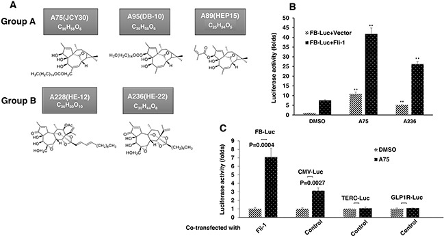
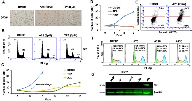
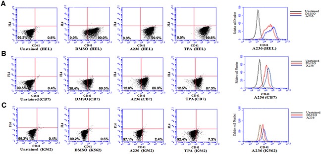
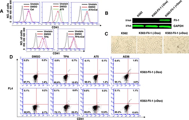
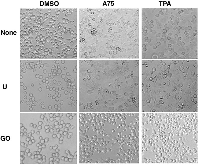
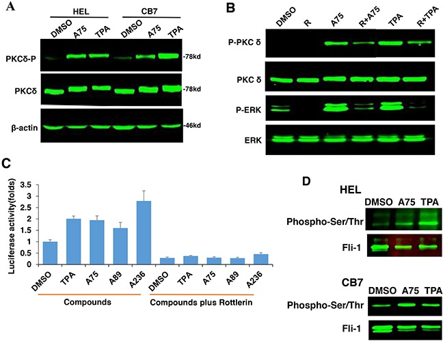
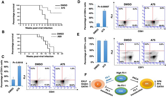
The ETS-related transcription factor Fli-1 affects many developmental programs including erythroid and megakaryocytic differentiation, and is frequently de-regulated in cancer. Fli-1 was initially isolated following retrovirus insertional mutagenesis screens for leukemic initiator genes, and accordingly, inhibition of this transcription factor can suppress leukemia through induction of erythroid differentiation. To search for modulators of Fli-1, we hereby performed repurposing drug screens with compounds isolated from Chinese medicinal plants. We identified agents that can transcriptionally activate or inhibit a Fli-1 reporter. Remarkably, agents that increased Fli-1 transcriptional activity conferred a strong anti-cancer activity upon Fli-1-expressing leukemic cells in culture. As opposed to drugs that suppress Fli1 activity and lead to erythroid differentiation, growth suppression by these new Fli-1 transactivating compounds involved erythroid to megakaryocytic conversion (EMC). The identified compounds are structurally related to diterpene family of small molecules, which are known agonists of protein kinase C (PKC). In accordance, these PKC agonists (PKCAs) induced PKC phosphorylation leading to activation of the mitogen-activated protein kinase (MAPK) pathway, increased cell attachment and EMC, whereas pharmacological inhibition of PKC or MAPK diminished the effect of our PKCAs. Moreover, in a mouse model of leukemia initiated by Fli-1 activation, the PKCA compounds exhibited strong anti-cancer activity, which was accompanied by increased presence of CD41/CD61 positive megakaryocytic cells in leukemic spleens. Thus, PKC agonists offer a novel approach to combat Fli-1-induced leukemia, and possibly other cancers,by inducing EMC in part through over-activation of the PKC-MAPK-Fli-1 pathway.

Keywords: Fli-1; PKC; drug screens; erythroid and megakaryocytic differentiation; leukemia therapy.


Conflict of interest statement

CONFLICTS OF INTEREST

The authors declare no conflicts of interest.

Figures

Figure 1
Figure 1. Identification of Fli-1 transcription activating compounds
A. Structure and chemical composition of Group A and B compounds. B. A75 and A236 compounds increase transcriptional activity of FB-Luc (1.25 μg) reporter gene co-transfected with MigR1-Fli-1 (1.25 μg) or MigR1 (1.25 μg) vectors into HEK293T cells. C. A75 and A236 compounds moderately increase luciferase activity of control CMV-Luc but not of TERC-Luc or GLP1R-Luc promoters. Compound concentration in panels B-C, 1μM. P values calculated by student t-test. P< 0.005 denoted by **.
Figure 2
Figure 2. Fli-1 transactivating compounds block proliferation, and induce polyploidy as well as attachment of Fli-1- expressing erythroleukemic cells in culture
A. Micrographs showingA75 and TPA increase the attachment and appearance of large multi-nuclear cells in CB7 cell line, 3 days post-drug incubation (magnification x40). B. CB7 cells treated with A75 or TPA exhibit increased polyploidy associated with appearance of 2N, 4N and 8N chromosomes, 72 hours post treatment. C. A75 and TPA strongly suppress growth of CB7 cells, even when the compounds are removed 3 days post drug treatment. D. A236 and TPA suppress growth of K562 cells in culture. E. A75 treatment increases the percentage of Annexin V-positive apoptotic cells in K562 cells 72 hours post drug treatment. F. A75, A228 and A236 increase G2/M and decrease S phase stages of cell cycle in K562 cells, 16 hour post-drug incubation. Compound Concentration in panels A-F, 5μM. G. Indicated compounds (2μM) did not induce Fli-1 expression in K562 cells, as determined by Western blot analysis. HEL cells were used as positive control.
Figure 3
Figure 3. Fli-1 transactivating compounds induce megakaryocytic differentiation in erythroleukemia cell lines
Treatment of the erythroleukemic cell lines HEL A. and CB7 B. with A236 and TPA increases the percentage and level of CD41 expression, 48 hours post- treatment. C. These compounds only weakly induce CD41 expression in K562 cells. Compound concentration, 5μM.
Figure 4
Figure 4. Induction of Fli-1 expression promotes megakaryocytic differentiation
A. The A75- and TPA- induced CD41 expression in HEL cells can be blocked by treatment with Calcimycin (5μM). B. Western blot of K562 and K562-Fli-1indu-20-on cells treated or not with Doxycycline (DOX) for, 24 hours. C. Induction of Fli-1 in K562-Fli-1 cells by DOX (1μM) increases the number of large, polynucleated cells compared to control cells. D. Induction of CD41 expressing cells in K562-Fli-1 cells with or without DOX treatment plus the indicated compounds.
Figure 5
Figure 5. The compounds induces Fli-1 transcriptional activity through MAPK/ERK
A. Indicated compounds induce phosphorylation of MAPK/ERK in CB7 cells. B. Induction of MAPK/ERK by A236 can be blocked by the MEK inhibitor U0126 (U). Phosphorylation was not blocked by JNK SP600125 (Sp) or P38 SB203580 (Sb) inhibitors, respectively, but partially by the RAF inhibitor Sorafenib tosylate (St). C. A75- and TPA-induced phosphorylation of MAPK/ERK is inhibited by U0126 in HEL cells. D. Induction of MAPK/ERK phosphorylation by A75 and TPA is blocked by addition of both U0126 and BCR-ABL inhibitor Imatinib in K562 cells, E. Fli-1 transcriptional activity induced by the indicated compounds in HEK293T cells co-transfected with FB-Luc+MigR1-Fli-1 is blocked by MEK inhibitor U0126. F. Western blot analysis for Fli-1 expression in CB7 and HEL cells after treatment with A75, A89 and A228. Compound concentration in experiments A-F, 2μM. Concentration of MAPK/ERK inhibitors, 5μM.
Figure 6
Figure 6. Fli-1 transactivating compounds alter expression of marker genes involved in erythroid to megakaryocytic differentiation
Expression of marker genes for erythroid and megakaryocytic lineages PF4 A., GP6 B., GATA1/2μM C., GATA1/5μM D., EKLF E., β-globin F. and RUNX1 G. in Fli-1 expressing HEL cells treated for 24 hours with TPA, A236 and A75, using RT-PCR (inserts) and Q-RT-PCR (graphs). Compound concentrations for experiments in panel A-F, 5μM. All Q-RT-PCRs were performed in triplicates and repeated at least 2 times. P< 0.05 denoted by *; P< 0.005 by **.
Figure 7
Figure 7. Induction of erythroid to megakaryocytic differentiation by compounds requires Fli-1
Expression of markers of erythroid and megakaryocytic lineages PF4 A., GP6 B., GATA1 C., EKLF D., β-globin E. and RUNX1 F. in Fli-1-negative K562 cells treated for 24 hours with or without TPA, A236 and A75 using RT-PCR (inserts) and Q-RT-PCR (graphs). Similar analysis in K562-Fli-1 cells with or without doxycycline (DOX) treatment for PF4 G., GP6 H., GATA1 I., EKLF J., β-globin K. and RUNX1 L., as indicated. Compound concentration, 5μM. Since PF4 expression is negligible in K562, HEL cells were used as a positive control (Panel A). All Q-RT-PCRs were performed in triplicates and repeated at least 2 times. P< 0.05 denoted by *; P< 0.005 by **.
Figure 8
Figure 8. The Fli-1 transactivating compounds induce growth arrest and differentiation of erythroleukemic cells through PKC and, in part, through MAPK/ERK activation
A. HEK293T cells co-transfected with FB-Luc and MigR1-Fli-1, and treated with A75 or TPA (1μM), show induction of Fli-1 transcriptional activity that can be blocked by U1026 (5μM) or GO6982 (2μM). B. Treatment of HEL or CB7 cells with A75 or TPA (2μM) induces MAPK/ERK phosphorylation that can be suppressed by GO6982 (2μM). C. Growth suppression induced by A75 or TPA (2μM) in HEL cells is reversed by GO6983 (2μM). D. Growth suppression induced in CB7 cells by A75 (2μM) is only marginally inhibited by U1026 (10μM). E, F. In CB7 cells, induction of megakaryocytic marker PF4 by A75 is completely inhibited by U0126 and GO6983 (5μM); these inhibitors suppressed downregulation of erythroid β-globin by A75. G. A model depicting the sequential events that occur in leukemic cells after treatment with PKCA compounds leading to Fli-1 transcriptional activation. PKCA compounds activate Fli-1 and also increase cell attachment independently of MAPK/ERK activation. PKCA compounds likely activate other pathways that further increase their growth inhibitory activity.
Figure 9
Figure 9. PKCA compounds induce cell attachment independently of MAPK/ERK activation
CB7 cells were cultured in the presence of A75 and TPA compounds (2μM) with or without MEK U0126 (5μM) or PKC inhibitors GO6983 (2μM), respectively, as indicated. Cells were photographed 24 hours after treatments (magnification x20).
Figure 10
Figure 10. PKCA-induced phosphorylation of PKCδ, MAPK/ERK and induction of Fli-1 transcriptional activation can be reversed by the PKCδ-inhibitor Rottlerin
A. A75 or TPA treatment (2μM) for 24 hours induces phosphorylation of PKCδ in both HEL and CB7 cell lines. B. Phosphorylation of MAPK/ERK and PKCδ in HEL cells treated with indicated compounds with or without PKCδ inhibitor Rottlerin. C. Fli-1 transcriptional activity induced by A75 and TPA compounds in HEK293T cells co-transfected with FB-Luc+MigR1-Fli-1 is blocked by Rottlerin. D. Extracts from CB7 and HEL cells were immunoprecipitated using anti-Fli-1 antibody followed by immune-blotting with anti-phospho-Serine/Threonine or anti-Fli-1 antibodies.
Figure 11
Figure 11. Distinct PKCA compounds can inhibit leukemogenesis in a mouse model of leukemia induced by Fli-1 retroviral insertional activation
A, B. Groups of BALB/c mice (n=7) were infected at birth with F-MuLV and 5 weeks later treated with A75 or A89 compounds (3 mg/ kg body weight), every other day for a total of six injections. Latency to death was used to plot a Kaplan-Meire survival curve. For the A75-treated group (A), the experiment was terminated around 20 week post-viral injection. Splenocytes isolated from large spleens of leukemic mice were used to determine the percentage of cells expressing megakaryocytic CD41 C. and CD61 D., or erythroid CD71 E. markers, using flow cytometry. Individual and average flow charts for each experiment is shown. F. A model for PKCA drug-induced erythroid-to-megakaryocytic differentiation in erythroleukemia. In HEL cells, Fli-1 downregulation (Fli-1 down) through either siRNA [6] or pharmacological drugs [23] leads to erythroid differentiation. Conversely, Fli-1 activation by these compounds promotes megakaryocytic differentiation. In K562 cells which express no Fli1, Fli-1 transduction (Fli-1 up) also induces megakaryocytic differentiation. This model suggests that both Fli-1 inactivation and activation suppress leukemia either through erythroid or megakaryocytic differentiation, respectively. E - erythrocytes; MK - megakaryocytes.

Similar articles

Cited by

References

    1. Johnston SJ, Carroll JS. Transcription factors and chromatin proteins as therapeutic targets in cancer. Biochim Biophys Acta. 2015;1855:183–92. - PubMed
    1. Li Y, Luo H, Liu T, Zacksenhaus E, Ben-David Y. The ets transcription factor Fli-1 in development, cancer and disease. Oncogene. 2015;34:2022–31. - PMC - PubMed
    1. Ben-David Y, Giddens EB, Bernstein A. Identification and mapping of a common proviral integration site Fli-1 in erythroleukemia cells induced by Friend murine leukemia virus. Proc Natl Acad Sci U S A. 1990;87:1332–6. - PMC - PubMed
    1. Ben-David Y, Giddens EB, Letwin K, Bernstein A. Erythroleukemia induction by Friend murine leukemia virus: insertional activation of a new member of the ets gene family, Fli-1, closely linked to c-ets-1. Genes Dev. 1991;5:908–18. - PubMed
    1. Delattre O, Zucman J, Plougastel B, Desmaze C, Melot T, Peter M, Kovar H, Joubert I, de Jong P, Rouleau G, Et A. Gene fusion with an ETS DNA-binding domain caused by chromosome translocation in human tumours. Nature. 1992;359:162–5. - PubMed

MeSH terms