. 2017 Feb 7;8(6):10114-10135.
doi: 10.18632/oncotarget.14357.

BRCA1-IRIS overexpression promotes and maintains the tumor initiating phenotype: implications for triple negative breast cancer early lesions

Affiliations

BRCA1-IRIS overexpression promotes and maintains the tumor initiating phenotype: implications for triple negative breast cancer early lesions

Abhilasha Sinha et al. Oncotarget. .

Abstract

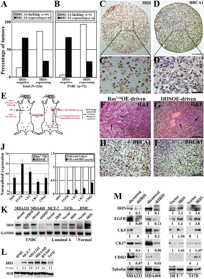
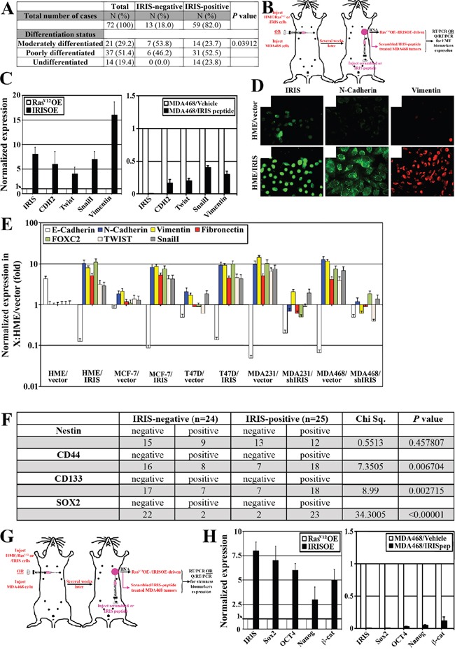
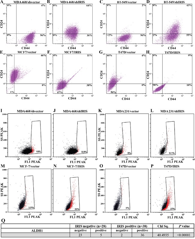
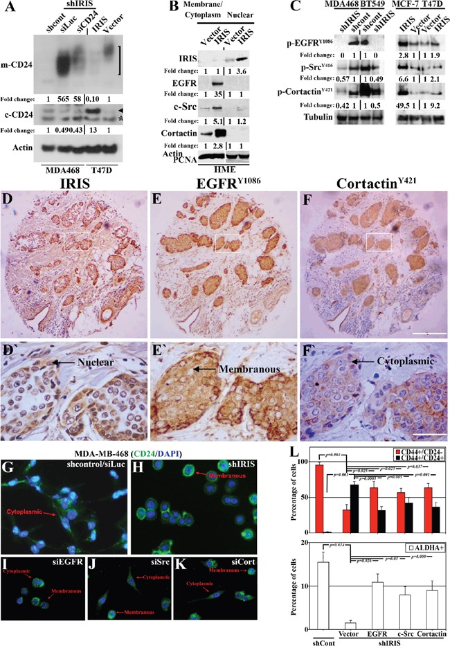
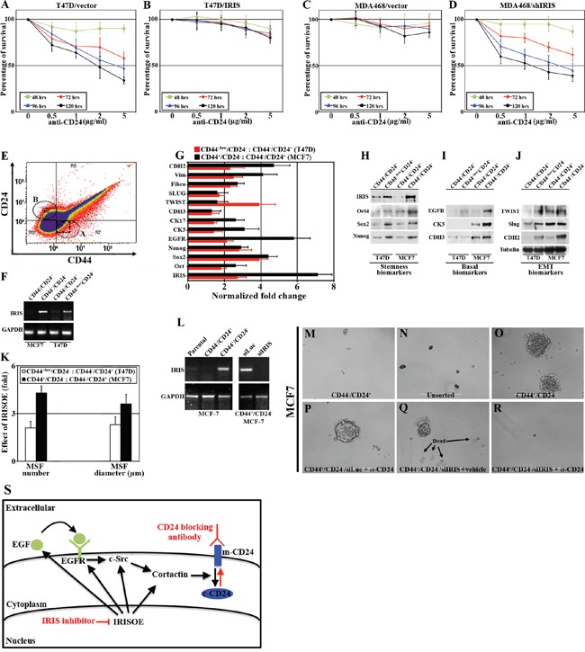
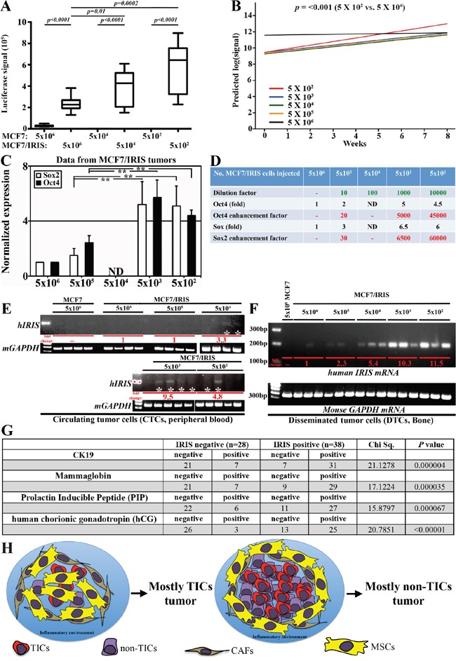
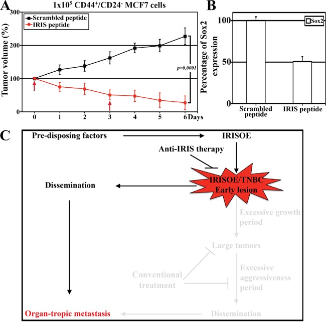
Tumor-initiating cells (TICs) are cancer cells endowed with self-renewal, multi-lineage differentiation, increased chemo-resistance, and in breast cancers the CD44+/CD24-/ALDH1+ phenotype. Triple negative breast cancers show lack of BRCA1 expression in addition to enhanced basal, epithelial-to-mesenchymal transition (EMT), and TIC phenotypes. BRCA1-IRIS (hereafter IRIS) is an oncogene produced by the alternative usage of the BRCA1 locus. IRIS is involved in induction of replication, transcription of selected oncogenes, and promoting breast cancer cells aggressiveness. Here, we demonstrate that IRIS overexpression (IRISOE) promotes TNBCs through suppressing BRCA1 expression, enhancing basal-biomarkers, EMT-inducers, and stemness-enforcers expression. IRISOE also activates the TIC phenotype in TNBC cells through elevating CD44 and ALDH1 expression/activity and preventing CD24 surface presentation by activating the internalization pathway EGFR→c-Src→cortactin. We show that the intrinsic sensitivity to an anti-CD24 cross-linking antibody-induced cell death in membranous CD24 expressing/luminal A cells could be acquired in cytoplasmic CD24 expressing IRISOE TNBC/TIC cells through IRIS silencing or inactivation. We show that fewer IRISOE TNBC/TICs cells form large tumors composed of TICs, resembling TNBCs early lesions in patients that contain metastatic precursors capable of disseminating and metastasizing at an early stage of the disease. IRIS-inhibitory peptide killed these IRISOE TNBC/TICs, in vivo and prevented their dissemination and metastasis. We propose IRIS inactivation could be pursued to prevent dissemination and metastasis from early TNBC tumor lesions in patients.

Keywords: BRCA1-IRIS; TNBC; breast cancer; early lesion; metastasis.


Conflict of interest statement

CONFLICTS OF INTEREST

Authors declare no conflicts of interest.

Figures

Figure 1
Figure 1. IRISOE suppresses BRCA1 expression and enhances basal-biomarkers expression in breast cancer cells
Immunohistochemical analysis of IRIS and BRCA1 expression in a cohort of breast tumor (all subtypes, n=326, A), or a sub-cohort of TNBC tumors (n=72, B). Representative images of IRISOE (C, and larger magnification C`) associated with lack of BRCA1 expression (D, and larger magnification D`) in a TNBC tumor sample. Scale bars: 300μm in C and D, and 50μm in C` and D`. E. Schematic of the strategy used to generate RasV12OE-/IRISOE-driven or MDA468 + scrambled/MDA468 + IRIS inhibitory peptide orthotopic mammary tumors in SCID/Nu/Nu mice, followed by tumor and RNA isolation and basal-biomarkers expression analysis. H&E (F and G) and BRCA1 (H and I) staining on RasV12-driven or IRISOE-driven orthotopic mammary tumors, respectively generated as in (E). Scale bars: 200μm in F and G, and 100μm in H and I. J. real-time QRT/PCR analysis for the expression of IRIS and several basal-biomarkers mRNA in RasV12OE-driven or IRISOE-driven orthotopic mammary tumors (left), and MDA468 orthotopic mammary tumors after treatment with scrambled- or IRIS-inhibitory peptide (right). K. RT/PCR analysis of IRIS mRNA in naïve HME or the luminal cell lines; MCF7 and T47D before and after IRISOE and the TNBC cell lines; MDA231 and MDA468 before and after IRIS knockdown. L. Comparison of IRIS protein expression in the luminal A cell lines; MCF7, T47D, and the TNBC cell lines; MDA231, MDA468 and BT-549 compared to naïve HME cells. M. Western blot analysis for the expression of several basal-biomarkers in the TNBC cell lines; MDA231 and MDA468 before and after IRIS-silencing, and the luminal cell lines; MCF7 and T47D before and after IRISOE.
Figure 2
Figure 2. IRISOE promotes expression of EMT-inducers and stemness-enforcers in breast cancers
A. Association analysis between IRIS expression and the differentiation status of TNBC tumors (n=72). B. Schematic of the strategy used to generate RasV12OE-/IRISOE-driven or MDA468 + scrambled/MDA468 + IRIS inhibitory peptide orthotopic mammary tumors in SCID/Nu/Nu mice, followed by tumor and RNA isolation and EMT-inducers expression analysis. C. Real-time QRT/PCR analysis showing the expression of IRIS and several EMT-inducers mRNAs in RasV12OE-driven or IRISOE-driven orthotopic mammary tumors (left), and MDA468 orthotopic mammary tumors after treatment with scrambled- or IRIS-inhibitory peptide (right), generated as in (B). D. Immunofluorescence analysis of IRIS, N-cadherin and vimentin expression in naïve (uppers) or IRISOE (lowers) HME cells. E. Real time QRT/PCR of indicated mRNAs in naïve/IRISOE HME cells, vector/IRIS transfected MCF7 or T47D, and shcontrol/shIRIS transfected MDA231 or MDA468. F. Association analysis between IRISOE and the expression of several TIC-biomarkers in a cohort of locally advanced breast cancer patients (n=49, from Egyptian NCI) conducted using real-time QRT/PCR. G. Schematic of the strategy used to generate RasV12OE-/IRISOE-driven or MDA468 + scrambled/MDA468 + IRIS inhibitory peptide orthotopic mammary tumors in SCID/Nu/Nu mice, followed by tumor and RNA isolation and stemness-enforcers expression analysis. H. Real-time QRT/PCR analysis for the expression of IRIS and the stemness-enforcers mRNAs expression in RasV12OE-driven or IRISOE-driven orthotopic mammary tumors (left), and MDA468 orthotopic mammary tumors after treatment with scrambled- or IRIS-inhibitory peptide (right), generated as (G).
Figure 3
Figure 3. IRISOE drives the TIC phenotype
FACS analysis for surface CD44 and CD24 double-staining (for 1h) populations within shcontrol- A., or shIRIS- B. expressing MDA468 cells, shcontrol- C., or shIRIS- D. expressing BT-549 cells, vector E., or IRIS- F. overexpressing MCF7, vector- G., or IRIS- H. overexpressing T47D cells. FACS analysis showing ALDH1+ (red) compared to ALDHA- (black) populations in shcontrol- I., or shIRIS- J. expressing MDA468 cells, shcontrol- K., or shIRIS- L. expressing MDA231 cells, vector M., or IRIS- N. overexpressing MCF7, vector- O., or IRIS- P. overexpressing T47D cells. Q. Association analysis of IRIS and the expression of ALDH1 in a cohort of metastatic breast cancer patients (n=66, from Egyptian NCI) conducted using real-time QRT/PCR.
Figure 4
Figure 4. Potential signaling pathway promotes CD24 cytoplasmic confinement in IRISOE TNBC cells
A. Western blot analysis for m-CD24 (open bracket, upper) or c-CD24 (arrowhead, lower, note asterisk indicates a non-specific band) in shcontrol- or shIRIS-expressing MDA468 cells expressing or not siCD24 (left), as well as vector or IRISOE T47D cells (right). B. Western blot analysis for the expression of nuclear IRIS and cytoplasmic/membranous EGFR, c-Src, and cortactin in naïve or IRISOE HME cells. C. Western blot analysis for the expression of p-EGFRY1086, p-SrcY416 and p-cortactinY421 in sonicates from shcontrol- or shIRIS-expressing MDA468 and BT-549 cells (left) and parental or IRIS-expressing MCF7 and T47D cells (right). The expression of IRIS D and D`. EGFRY1086 E and E`. and cortactinY421 F and F`. detected using IHC in a TNBC tumor sample. G-K. Immunofluorescence analysis for the expression of CD24 in shcontrol- (G), shIRIS-expressing (H), transiently siEGFR (I), siSrc (J), or sicortactin (K) transfected MDA468 cells. L. FACS analysis showing the percentage of CD44+/CD24- and CD44+/CD24+ (upper) and ALDHA+ (lower) in shcontrol-, or shIRIS-expressing MDA468 cells without and with EGFR, Src, or cortactin transient overexpression.
Figure 5
Figure 5. Intrinsic sensitivity in luminal A cancer cells to an anti-CD24 blocking antibody could be acquired in TNBC/TIC cells following IRIS inhibition
Percentage of survival in vector- A. or IRIS- B. overexpressing T47D cells, as well as shcontrol- C. or shIRIS- D. expressing MDA468 cells following incubation with increasing concentration of anti-CD24 monoclonal antibody for the indicated times. E. FACS analysis of non-permeabilized MCF7 cells incubated with FITC-CD44 and PE-CD24 antibodies for only 20mins. F. Expression of IRIS mRNA in non-CD44-/CD24+ and TIC-CD44+/CD24- MCF7 cells, or non-CD44-/CD24+ and TIC-like-CD44-/CD24- T47D cells sorted as in (E). G. Real time QRT/PCR analysis for the expression of IRIS and the indicated basal-biomarkers, EMT-inducers, and stemness-enforcers mRNA in MCF7 TIC-CD44+/CD24- cells compared to non-CD44-/CD24+ cells, and T47D TIC-like-CD44-/CD24- cells compared to non-CD44-/CD24+ cells sorted as in (E). H-J. Western blot analysis for the expression of IRIS and the indicated basal-biomarkers, EMT-inducers, and stemness-enforcers non-CD44-/CD24+ and TIC-CD44+/CD24- MCF7 cells, or non-CD44-/CD24+ and TIC-like-CD44-/CD24- T47D cells sorted as in (E). K. Numbers and diameters of mammospheres developed by non-CD44-/CD24+ and TIC-CD44+/CD24- MCF7 cells, or non-CD44-/CD24+ and TIC-like-CD44-/CD24- T47D cells sorted as in (E). L. RT/PCR analysis of IRIS mRNA in parental, non-TIC and TIC MCF7 cells sorted as in (E) before and after IRIS silencing. Mammosphere formation by non-TIC M. parental unsorted N., TIC O. MCF7 cells before or after anti-CD24 blocking antibody + siLuc P. vehicle + siIRIS Q. or anti-CD24 blocking antibody + siIRIS R. treatment. S. Schematic presentation of our hypothesis of sensitizing TNBC/TIC cells to anti-CD24 blocking antibody through IRIS silencing/inhibition.
Figure 6
Figure 6. IRISOE promotes formation of TIC tumors, their dissemination and metastasis
A. Luciferase signals obtained in tumors developed using different concentration of parental or IRISOE MCF7 at week 8 post cells injection. B. Slopes of curves generated for luciferase signals for tumors developed using MCF7/IRISOE cells originally injected at the indicted numbers plotted against time (weeks). C. Real time QRT/PCR analysis for human Sox2 and Oct4 mRNAs expression in tumors developed using MCF7/IRISOE cells originally injected at the indicated numbers at week 8. D. Assessment of the stemness enhancement factor in tumors developed using MCF7/IRISOE originally injected at the indicated numbers extrapolated from the data presented in (C). RT/PCR analysis for human IRIS mRNA (upper) or mouse GAPDH mRNA (loading control, lower) in PB E. or BM F. samples from mice originally injected with the indicated cell lines at the indicated numbers. G. Association between IRIS mRNA expression and the expression of the dissemination biomarkers; CK19, MGA, PIP, hGC mRNAs in a cohort of metastatic breast cancer patients (n=66, from Egyptian NCI) conducted using real-time QRT/PCR. H. Schematic presentation of our overall hypothesis that bidirectional interactions with the microenvironment enhance IRISOE/TNBC/TIC early lesion formation, aggressiveness, and eventually dissemination and metastasis.
Figure 7
Figure 7. The efficacy of IRIS inhibitory peptide against TIC tumor cells, in vivo
A. The growth of established TIC/CD44+CD- MCF7 tumors grown in NSG mice following scrambled or IRIS-inhibitory intratumoral injection at the indicated times (arrows). B. Real-time QRT/PCR analysis for the expression of human Sox2 mRNA in the tumors found in the mice at day 7 (in A) post-injection of scrambled or IRIS-inhibitory peptide. C. Schematic presentation of our data combined and the hypothesis that finding and targeting the early lesions could be much more beneficial for IRISOE/TNBC patients.

References

    1. Lerma E, Barnadas A, Prat J. Triple negative breast carcinomas: similarities and differences with basal like carcinomas. Appl Immunohistochem Mol Morphol. 2009;17:483–94. doi: 10.1097/PAI.0b013e3181a725eb. - DOI - PubMed
    1. Palma G, Frasci G, Chirico A, Esposito E, Siani C, Saturnino C, Arra C, Ciliberto G, Giordano A, D’Aiuto M. Triple negative breast cancer: looking for the missing link between biology and treatments. Oncotarget. 2015;6:26560–74. doi: 10.18632/oncotarget.5306. - DOI - PMC - PubMed
    1. Brady-West DC, McGrowder DA. Triple negative breast cancer: therapeutic and prognostic implications. Asian Pac J Cancer Prev. 2011;12:2139–43. - PubMed
    1. Korsching E, Jeffrey SS, Meinerz W, Decker T, Boecker W, Buerger H. Basal carcinoma of the breast revisited: an old entity with new interpretations. J Clin Pathol. 2008;61:553–60. doi: 10.1136/jcp.2008.055475. - DOI - PubMed
    1. Kallel I, Khabir A, Boujelbene N, Abdennadher R, Daoud J, Frikha M, Aifa S, Sallemi-Boudawara T, Rebaï A. EGFR overexpression relates to triple negative profile and poor prognosis in breast cancer patients in Tunisia. J Recept Signal Transduct Res. 2012;32:142–49. doi: 10.3109/10799893.2012.664552. - DOI - PubMed

MeSH terms

LinkOut - more resources