. 2017 Feb 7;8(6):10145-10160.
doi: 10.18632/oncotarget.14358.

Angiomotin regulates prostate cancer cell proliferation by signaling through the Hippo-YAP pathway

Affiliations

Angiomotin regulates prostate cancer cell proliferation by signaling through the Hippo-YAP pathway

Hao Zeng et al. Oncotarget. .

Abstract

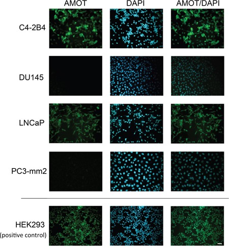
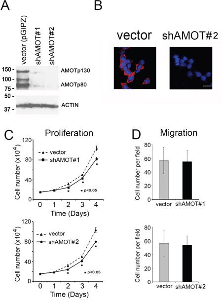
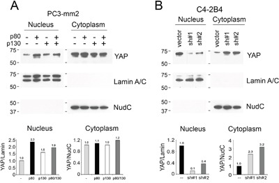
Angiomotin (AMOT) is a family of proteins found to be a component of the apical junctional complex of vertebrate epithelial cells and is recently found to play important roles in neurofibromatosis type 2 (NF-2). Whether AMOT plays a role in prostate cancer (PCa) is unknown. AMOT is expressed as two isoforms, AMOTp80 and AMOTp130, which has a 409 aa N-terminal domain that is absent in AMOTp80. Both AMOTp80 and AMOTp130 are expressed in LNCaP and C4-2B4, but at a low to undetectable level in PC3, DU145, and BPH1 cells. Further study showed that AMOTp130 and AMOTp80 have distinct functions in PCa cells. We found that AMOTp80, but not AMOT p130, functioned as a tumor promoter by enhancing PCa cell proliferation. Mechanistic studies showed that AMOTp80 signaled through the Hippo pathway by promoting nuclear translocation of YAP, resulting in an increased expression of YAP target protein BMP4. Moreover, inhibition of BMP receptor activity by LDN-193189 abrogates AMOTp80-mediated cell proliferation. Together, this study reveals a novel mechanism whereby the AMOTp80-Merlin-MST1-LATS-YAP-BMP4 pathway leads to AMOTp80-induced tumor cell proliferation.

Keywords: BMP4; Hippo pathway; YAP; angiomotin; proliferation.


Conflict of interest statement

CONFLICTS OF INTEREST

The authors declare no conflicts of interest.

Figures

Figure 1
Figure 1. Expression of AMOT isoforms in PCa cell lines
A. Left panel, Diagram of AMOTp80 and AMOTp130 proteins. Right panel, AMOTp80 and AMOTp130 messages were detected by real-time PCR using isoform-specific primers. B. Specificity of anti-AMOT antibody. PC3-mm2 cells stably expressing 7 histidine-tagged AMOTp80, AMOTp130, or vector alone were immunoblotted with anti-AMOT or anti-His antibody. HEK293 cells were used as a control for endogenous AMOT expression. C. Expression of AMOT isoforms in PCa cell lines. Equal amounts of cell lysates from PCa cell lines were immunoblotted with anti-AMOT antibody. Actin was used as a loading control. D. Subcutaneous tumors were generated from C4-2B4 and PC3 PCa cells. Formalin-fixed paraffin-embedded PCa tumors were immunostained with anti-AMOT antibody and the signals detected by DAB staining.
Figure 2
Figure 2. Expression of Amot in PCa cell lines
PCa cells on cover slips were immunostained with anti-AMOT antibody and counterstained with DAPI. HEK293 cells were used as a positive control. Scale bar, 50 μm.
Figure 3
Figure 3. Effect of AMOT on the proliferation and migration of PC3-mm2 cells
PC3-mm2 cells overexpressed A. AMOTp80, B. AMOTp130 or C. AMOTp80/p130 were analyzed for protein expression (left) by using anti-AMOT antibody, proliferation (middle) by using cell count, and migration (right) by using Boyden chamber assay. D. Proliferation of PC3-mm2 cells overexpressing AMOTp80, AMOTp130, or both was performed in the same experiment for a direct comparison. E. Overexpression of AMOTp80, AMOTp130, and both AMOTp80/p130 was analyzed by immunofluorescence. Scale bar, 20 μm
Figure 4
Figure 4. Effect of AMOT knockdown on the proliferation and migration of C4-2B4 cells
A. Western blot of cell lysates from C4-2B4 cells transduced with pGIPZ control vector, shAMOT RNA #1 or #2 vectors using anti-AMOT antibody. B. Knock down of AMOT by using shRNA#2 was analyzed by immunofluorescence. Scale bar, 20 μm C. Proliferation of C4-2B4 cells with AMOT knockdown. D. Migration of C4-2B4 cells with AMOT knockdown.
Figure 5
Figure 5. Effect of AMOT on the Hippo-YAP signaling pathway in PC3-mm2 cells
A. Left panel, PC3-mm2 cells overexpressing AMOTp80, AMOTp130, or both AMOTp80/p130 were cultured under normal serum condition. The levels of pYAP, YAP, pLATS, LATS, MST1 and MST2 were examined by western blot. Right panel, the relative expression of these proteins after normalization against actin. B. To examine the influence of serum, PC3-mm2 cells overexpressing AMOTp80, AMOTp130, or both AMOTp80/p130 were cultured overnight under serum starvation condition followed with serum stimulation. Western blot was performed and normalized as described in (A).
Figure 6
Figure 6. Effect of AMOT on the Hippo-YAP signaling pathway in C4-2B4 cells
C4-2B4 cells were transduced with pGIPZ control vector, AMOT shRNA #1, or AMOT shRNA#2 in pGIPZ vectors under normal serum condition or serum starvation overnight followed with serum stimulation condition. Western blot was used to detect the expression levels of the proteins and changes in their phosphorylation after normalization against actin.
Figure 7
Figure 7. Effect of AMOT on the cellular localization of YAP
Nuclear and cytoplasmic fractionation of A. PC3-mm2 cells overexpressing AMOTp80, AMOTp130, or both; or B. C4-2B4 cells with AMOT knockdown. The relative levels of YAP in the nucleus and cytoplasm were normalized to Lamin A/C, a protein found in the nucleus, or NudC, a protein found in the cytoplasm, respectively.
Figure 8
Figure 8. Effect of AMOT on YAP target gene expression and the involvement of BMP4 in AMOT-mediated PCa cell proliferation
A. PC-3mm2 cells overexpressing AMOTp80, AMOTp130, or both, and C4-2B4 cells with knock-down of AMOT by pGIPZ control vector, shAMOT RNA #1 or #2 in pGIPZ vectors. YAP target genes were detected by real-time PCR using specific primers. B. Left panel, PC3-mm2 cells overexpressing AMOTp80, p130, or both were incubated with an increasing concentration of LDN193189, a BMP Type I receptor antagonist, for two days and the number of cells was counted. Right panel, PC3-mm2 cells overexpressing AMOTp80, p130, or both were treated with 500 nM LDN193189. C. Left panel, C4-2B4 cells with AMOT knockdown were treated with an increasing concentration of LDN193189 and the number of cells was counted. Right panel, C4-2B4 cells with AMOT knockdown were treated with 200 nM LDN193189. D. Working model of AMOTp80-mediated PCa cell proliferation. AMOTp80 likely interacts with Merlin and this leads to decreases in the levels of MST1, pLATS and pYAP. The ensuing decrease in YAP phosphorylation leads to an increase in the nuclear translocation of YAP. YAP activation stimulates the transcription of BMP4, resulting in an increase in PCa cell proliferation.

References

    1. Ernkvist M, Luna Persson N, Audebert S, Lecine P, Sinha I, Liu M, Schlueter M, Horowitz A, Aase K, Weide T, Borg JP, Majumdar A, Holmgren L. The Amot/Patj/Syx signaling complex spatially controls RhoA GTPase activity in migrating endothelial cells. Blood. 2009;113:244–253. - PMC - PubMed
    1. Troyanovsky B, Levchenko T, Mansson G, Matvijenko O, Holmgren L. Angiomotin: an angiostatin binding protein that regulates endothelial cell migration and tube formation. J Cell Biol. 2001;152:1247–1254. - PMC - PubMed
    1. Wells CD, Fawcett JP, Traweger A, Yamanaka Y, Goudreault M, Elder K, Kulkarni S, Gish G, Virag C, Lim C, Colwill K, Starostine A, et al. Pawson T. A Rich1/Amot complex regulates the Cdc42 GTPase and apical-polarity proteins in epithelial cells. Cell. 2006;125:535–548. - PubMed
    1. Sugihara-Mizuno Y, Adachi M, Kobayashi Y, Hamazaki Y, Nishimura M, Imai T, Furuse M, Tsukita S. Molecular characterization of angiomotin/JEAP family proteins: interaction with MUPP1/Patj and their endogenous properties. Genes Cells. 2007;12:473–486. - PubMed
    1. Yi C, Troutman S, Fera D, Stemmer-Rachamimov A, Avila JL, Christian N, Persson NL, Shimono A, Speicher DW, Marmorstein R, Holmgren L, Kissil JL. A tight junction-associated Merlin-angiomotin complex mediates Merlin's regulation of mitogenic signaling and tumor suppressive functions. Cancer Cell. 2011;19:527–540. - PMC - PubMed

MeSH terms