. 2017 Jan 4;13(1):e1006146.
doi: 10.1371/journal.ppat.1006146. eCollection 2017 Jan.

CRL4WDR1 Controls Polo-like Kinase Protein Abundance to Promote Bilobe Duplication, Basal Body Segregation and Flagellum Attachment in Trypanosoma brucei

Affiliations

CRL4WDR1 Controls Polo-like Kinase Protein Abundance to Promote Bilobe Duplication, Basal Body Segregation and Flagellum Attachment in Trypanosoma brucei

Huiqing Hu et al. PLoS Pathog. .

Abstract

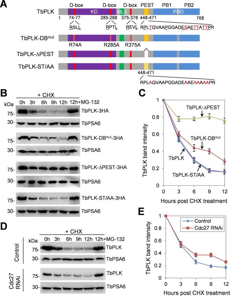
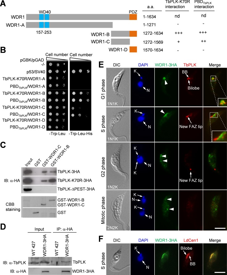
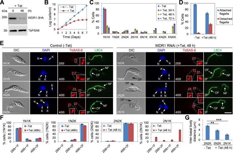
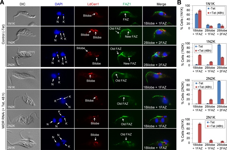
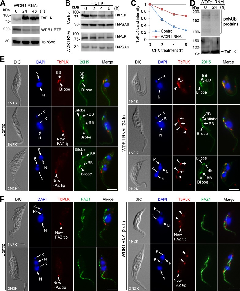
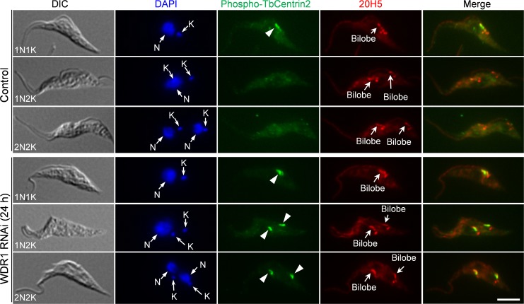
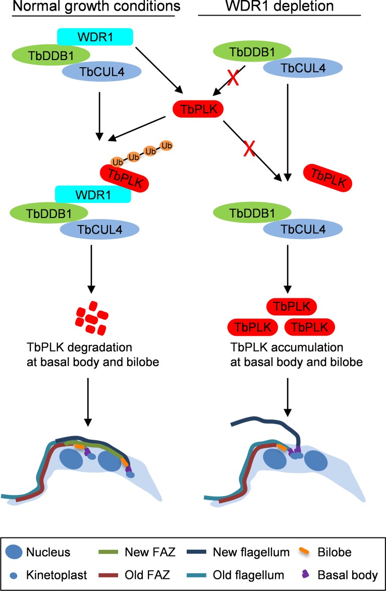
The Polo-like kinase homolog in Trypanosoma brucei, TbPLK, plays essential roles in basal body segregation, flagellum attachment and cytokinesis. The level of TbPLK protein is tightly controlled, but the underlying mechanism remains elusive. Here, we report a Cullin-RING ubiquitin ligase composed of Cullin4, the DNA damage-binding protein 1 homolog TbDDB1 and a WD40-repeat protein WDR1 that controls TbPLK abundance in the basal body and the bilobe. WDR1, through its C-terminal domain, interacts with the PEST motif in TbPLK and, through its N-terminal WD40 motif, binds to TbDDB1. Depletion of WDR1 inhibits bilobe duplication and basal body segregation, disrupts the assembly of the new flagellum attachment zone filament and detaches the new flagellum. Consistent with its role in TbPLK degradation, depletion of WDR1 causes excessive accumulation of TbPLK in the basal body and the bilobe, leading to continuous phosphorylation of TbCentrin2 in the bilobe at late cell cycle stages. Together, these results identify a novel WD40-repeat protein as a TbPLK receptor in the Cullin4-DDB1 ubiquitin ligase complex for degrading TbPLK in the basal body and the bilobe after the G1/S cell cycle transition, thereby promoting bilobe duplication, basal body separation and flagellum-cell body adhesion.


Conflict of interest statement

We declare no competing interests.

Figures

Fig 1
Fig 1. Degradation of TbPLK is mediated by destruction box and PEST motif.
(A). Schematic drawing of the domains in TbPLK and the various mutants of TbPLK expressed for protein stability assay. DB, destruction box or D-box; KD, kinase domain; NLS, nuclear localization signal sequence; PEST, proline (P), glutamic acid (E), serine (S) and threonine (T)-enriched sequence; PBD, Polo-box domain; PB1 and PB2, Polo-boxes 1 and 2. The signature arginine (R) and leucine (L) residues in the three D-boxes, and the proline (P), glutamate (E), serine (S) and threonine (T) residues in the PEST motif are underlined. The phosphorylated serine and threonine residues in the PEST motif are highlighted in red. (B). Degradation of ectopically overexpressed TbPLK and its D-box mutant and PEST-deletion mutant. Cells overexpressing 3HA-tagged TbPLK or each of the TbPLK mutants were treated with cycloheximide, and time-course samples (equal numbers of cells) were collected for Western blotting with anti-HA antibody. In a separate cell sample, MG-132 was added together with cycloheximide and incubated for 12 h (12h+MG-132). TbPSA6, the T. brucei proteasome subunit alpha-6, served as the loading control. (C). Quantification of TbPLK band intensity from panel B. TbPLK band intensity was measured with ImageJ, and normalized with the band intensity of TbPSA6. Error bars represent S.D. calculated from three independent experiments. (D). Degradation of TbPLK in control and Cdc27 RNAi cells, which was induced for 72 h. Cells were treated with cycloheximide for up to 12 h, and time-course samples were collected for Western blotting with anti-TbPLK antibody. The level of TbPSA6 served as the loading control. (E). Quantification of TbPLK band intensity from panel D. TbPLK band intensity was measured with ImageJ, and normalized with the band intensity of TbPSA6. Error bars indicate S.D. calculated from three independent experiments.
Fig 2
Fig 2. WDR1 interacts with TbPLK through its C-terminal domain and partially overlaps with TbPLK in the basal body and the bilobe at G1 phase.
(A). Schematic drawing of the domains in WDR1 and the truncation mutants of WDR1, and summary of yeast two-hybrid results. nd, not done because we failed to clone the full-length WDR1 gene into the yeast expression vectors despite multiple attempts. (B). Directional yeast two-hybrid assay to detect the interaction between TbPLK-K70R, PBDTbPLK and WDR1 truncation mutants. (C). WDR1, through its C-terminal domain, interacts with the PEST motif of TbPLK in vitro. The truncation fragments of WDR1 were expressed as GST-fusion proteins in E. coli, purified and used to pull down TbPLK-3HA, TbPLK-K70R-3HA, and TbPLK-ΔPEST-3HA from T. brucei cell lysate. CBB, coomassie brilliant blue. (D). WDR1 interacts with TbPLK in vivo in T. brucei, as demonstrated by co-immunoprecipitation. Endogenously 3HA-tagged WDR1 was immunoprecipitated with anti-HA antibody conjugated to protein G sepharose beads, and immunoprecipitated proteins were immunoblotted with anti-TbPLK antibody and anti-HA antibody to detect TbPLK and WDR1-3HA, respectively. (E). Subcellular localization of WDR1 and TbPLK during the cell cycle. Cells expressing endogenously 3HA-tagged WDR1 were co-immunostained with FITC-conjugated anti-HA mAb and anti-TbPLK pAb, and counterstained with DAPI for nuclear (N) and kinetoplast (K) DNA. Scale bar: 5 μm. (F). Localization of WDR1 to the basal body and the bilobe region during the S phase of the cell cycle. Cells were co-immunostained with FITC-conjugated anti-HA mAb to label WDR1-3HA and anti-LdCen1 pAb to label the basal body and the bilobe. Among the 115 S-phase cells examined, all of them showed WDR1-3HA localization to the basal body and the bilobe region. Scale bar: 5 μm.
Fig 3
Fig 3. WDR1 forms a complex with TbCUL4 and TbDDB1.
(A). Alignment of the putative DWD box in WDR1 with the DWD box of fission yeast and human WD40-repeat proteins that have been confirmed to bind to DDB1. The three highly conserved residues are highlighted in red, and other conserved residues are in green. The consensus sequence of the DWD box is shown at the top of the aligned sequences. Tb, T. brucei; Sp, Schizosaccharomyces pombe; Hs, Homo sapiens. (B). WDR1 interacts with TbCUL4 but not other Cullin proteins, in T. brucei, as demonstrated by co-immunoprecipitation. WDR1-3HA and each of the five PTP-tagged Cullin proteins were co-expressed from their respective endogenous locus in T. brucei. Immunoprecipitation was performed by incubating the cell lysate with IgG beads, and immunoprecipitated proteins were then immunoblotted with anti-HA antibody and anti-Protein A (α-ProtA) antibody, respectively. (C). WDR1 interacts with TbDDB1 in vivo in T. brucei, as demonstrated by co-immunoprecipitation. WDR1-3HA and TbDDB1-PTP were co-expressed in T. brucei, and immunoprecipitation and Western blotting were performed as described in panel B. (D). WDR1, TbCUL4, TbDDB1 and TbPLK form a complex in T. brucei, as demonstrated by co-immunoprecipitation. WDR1-3HA, TbCUL4-PTP and TbDDB1-3Myc were co-expressed from their respective endogenous locus in T. brucei. Immunoprecipitation of WDR1-3HA was carried out by incubating the cell lysate with EZview Red anti-HA affinity gel, and immunoprecipitated proteins were immunoblotted with anti-HA antibody, anti-Myc antibody, anti-TbPLK antibody and anti-Protein A (α-ProtA) antibody to detect WDR1-3HA, TbDDB1-3Myc, TbPLK and TbCUL4-PTP, respectively. (E). The N-terminal domain of WDR1 mediates the interaction with TbDDB1, as demonstrated by in vitro GST pull-down. The N-terminal fragment (1–300 aa) of WDR1, which contains the WD40 motif, was expressed as a GST-fusion protein in E. coli, purified and used to pull down TbDDB1-3HA from T. brucei cell lysate. Purified GST-WDR11-300 and GST were stained by coomassie brilliant blue (CBB).
Fig 4
Fig 4. WDR1 depletion inhibited basal body segregation and caused flagellum detachment.
(A). Western blotting to monitor the level of WDR1-3HA, which was endogenously tagged in the WDR1 RNAi cell line, before and after tetracycline induction of WDR1 RNAi. The level of TbPSA6 was included as the loading control. (B). RNAi of WDR1 caused a severe growth defect. Cells harboring the WDR1 RNAi construct were incubated with (+Tet) or without (-Tet) tetracycline and counted every day for 7 days. Shown is the result from one of the four clonal RNAi cell lines that target two different sequences of the WDR1 gene. (C). Quantification of cells with different numbers of nucleus (N) and kinetoplast (K) before and after WDR1 RNAi. 200 cells were counted for each time point. Error bars represent S.D. (D). Percentage of cells with attached or detached flagellum before (-Tet) and after (+Tet) WDR1 RNAi induction for 48 h. 200 cells were counted from each time point, and error bars represent S.D. (E). Effect of WDR1 depletion on basal body segregation and flagellum attachment. Cells were immunostained with anti-TbSAS-6 pAb and L8C4 (anti-PFR2 mAb) to label the basal body (BB; including mature BB and pro-BB) and the flagellum, respectively. Black arrows in the DIC channel indicate the detached flagellum (DF). Scare bar: 5 μm. (F). Quantification of basal body (BB) and flagellum in non-induced control (-Tet) and WDR1 RNAi cells (+Tet, 48 h). 200 cells of each cell type were counted, and error bars indicate S.D. (G). Inter-basal body distances of 2N2K and 2N1K cells before and after WDR1 RNAi. WDR1 RNAi was induced for 48 h, and non-induced control and WDR1 RNAi cells were immunostained with YL 1/2 and anti-TbSAS-6 antibodies. Distance between the two pairs of mature basal body/pro-basal body was measured with the ImageJ software. 200 cells of each cell type from control and WDR1 RNAi were counted, and error bars indicate S.D. from three independent experiments. Statistical analysis was performed using t-test in Excel. **, p<0.01; ***, p<0.001.
Fig 5
Fig 5. RNAi of WDR1 disrupted bilobe duplication and new FAZ filament assembly.
(A). Non-induced control cells (-Tet) and WDR1 RNAi-induced cells (+Tet, 48 h) were co-immunostained with L3B2, which labels the FAZ1 protein in the FAZ filament, and LdCen1, which detects TbCentrin4 in the bilobe, and counterstained with DAPI for nuclear (N) and kinetoplast (K) DNA. Scare bar: 5 μm. (B). Quantification of cells with different numbers of bilobe and FAZ filament in 1N1K, 1N2K, 2N2K and 2N1K cells. 300 cells were counted for each cell type at each time point, and error bars represent S.D.
Fig 6
Fig 6. Depletion of WDR1 caused excessive accumulation of TbPLK in the basal body and the bilobe.
(A). Western blotting to detect the level of TbPLK in control and WDR1 RNAi cells that were induced with tetracycline for 24 and 48 hours. TbPLK was detected by anti-TbPLK antibody, and WDR1 was endogenously tagged with a PTP epitope and detected with anti-Protein A antibody. TbPSA6 served as the loading control. Three repeats were performed, and all showed similar results. (B). Degradation of TbPLK in control and WDR1 RNAi cells. The non-induced control cells and WDR1 RNAi cells (24 h) were treated with cycloheximide for up to 6 h, and time course samples were collected at various times after cycloheximide treatment for Western blotting with anti-TbPLK antibody. TbPSA6 served as the loading control. (C). Quantification of TbPLK band intensity shown in panel B. TbPLK band intensity was determined with ImageJ, and normalized with the band intensity of TbPSA6. Error bars indicate S.D. calculated from three independent experiments. (D). Depletion of WDR1 reduced poly-ubiquitinated proteins of TbPLK immunoprecipitates. TbPLK was immunoprecipitated with anti-TbPLK pAb from non-induced control and WDR1 RNAi cells (24 h), and immunoprecipitated proteins were separated by SDS-PAGE and immunoblotted with anti-ubiquitin mAb and anti-TbPLK pAb to detect poly-ubiquitinated proteins (polyUB proteins) and TbPLK, respectively. Three repeats were performed, which showed similar results. (E, F). Effect of WDR1 depletion on TbPLK localization. WDR1 RNAi was induced for 24 h, and cells were co-immunostained with anti-TbPLK pAb and 20H5, which detects centrins in the basal body and the bilobe (panel E), and with anti-TbPLK pAb and anti-FAZ1 mAb, which labels the FAZ filament (Panel F). The arrows, solid arrowheads and open arrowheads in the TbPLK channel of panel E indicate the localization of TbPLK in the basal body, the bilobe and the new FAZ tip, respectively. The solid arrowheads in the TbPLK channel of panel F indicate the localization of TbPLK in the bilobe and the basal body region. N, nuclear DNA; K, kinetoplast DNA. Scale bars: 5 μm.
Fig 7
Fig 7. Effect of WDR1 depletion on TbPLK-mediated phosphorylation of TbCentrin2 in the bilobe.
Control and WDR1 RNAi cells (24 h) were co-immunostained with PS54 antibody, which detects the phospho-Ser54 of TbCentrin2 (arrowheads), and 20H5, which labels the bilobe. Cells were counterstained with DAPI for nuclear (N) and kinetoplast (K) DNA. Scale bar: 5 μm.
Fig 8
Fig 8. Model of the role of CRL4WDR1 in controlling TbPLK protein abundance to promote bilobe duplication, basal body segregation and FAZ assembly.
WDR1 in the CRL4 ubiquitin ligase complex recognizes the PEST motif in TbPLK. Binding of TbPLK to WDR1 causes TbPLK ubiquitination by CRL4WDR1 and subsequent degradation of TbPLK in the basal body and the bilobe after the G1/S cell cycle transition. This degradation of TbPLK in the basal body and the bilobe promotes bilobe duplication, basal body segregation, FAZ filament assembly, flagellum attachment and faithful cytokinesis. When WDR1 is depleted, TbPLK is not recruited to the TbCUL4-TbDDB1 complex for ubiquitination, leading to TbPLK accumulation in the basal body and the bilobe, where it may continuously phosphorylate the bilobe protein TbCentrin2. This accumulation of TbPLK inhibits bilobe duplication, impairs basal body segregation, disrupts the assembly of the new FAZ filament, which causes flagellum detachment, and blocks cytokinesis.

Similar articles

Cited by

References

    1. Barr FA, Sillje HH, Nigg EA (2004) Polo-like kinases and the orchestration of cell division. Nat Rev Mol Cell Biol 5: 429–440. 10.1038/nrm1401 - DOI - PubMed
    1. Archambault V, Glover DM (2009) Polo-like kinases: conservation and divergence in their functions and regulation. Nat Rev Mol Cell Biol 10: 265–275. 10.1038/nrm2653 - DOI - PubMed
    1. Lee KS, Grenfell TZ, Yarm FR, Erikson RL (1998) Mutation of the polo-box disrupts localization and mitotic functions of the mammalian polo kinase Plk. Proc Natl Acad Sci U S A 95: 9301–9306. - PMC - PubMed
    1. Song S, Grenfell TZ, Garfield S, Erikson RL, Lee KS (2000) Essential function of the polo box of Cdc5 in subcellular localization and induction of cytokinetic structures. Mol Cell Biol 20: 286–298. - PMC - PubMed
    1. Jang YJ, Lin CY, Ma S, Erikson RL (2002) Functional studies on the role of the C-terminal domain of mammalian polo-like kinase. Proc Natl Acad Sci U S A 99: 1984–1989. 10.1073/pnas.042689299 - DOI - PMC - PubMed

MeSH terms

LinkOut - more resources