. 2017 Jan 4;13(1):e1006148.
doi: 10.1371/journal.ppat.1006148. eCollection 2017 Jan.

Mapping Polyclonal HIV-1 Antibody Responses via Next-Generation Neutralization Fingerprinting

Affiliations

Mapping Polyclonal HIV-1 Antibody Responses via Next-Generation Neutralization Fingerprinting

Nicole A Doria-Rose et al. PLoS Pathog. .

Abstract

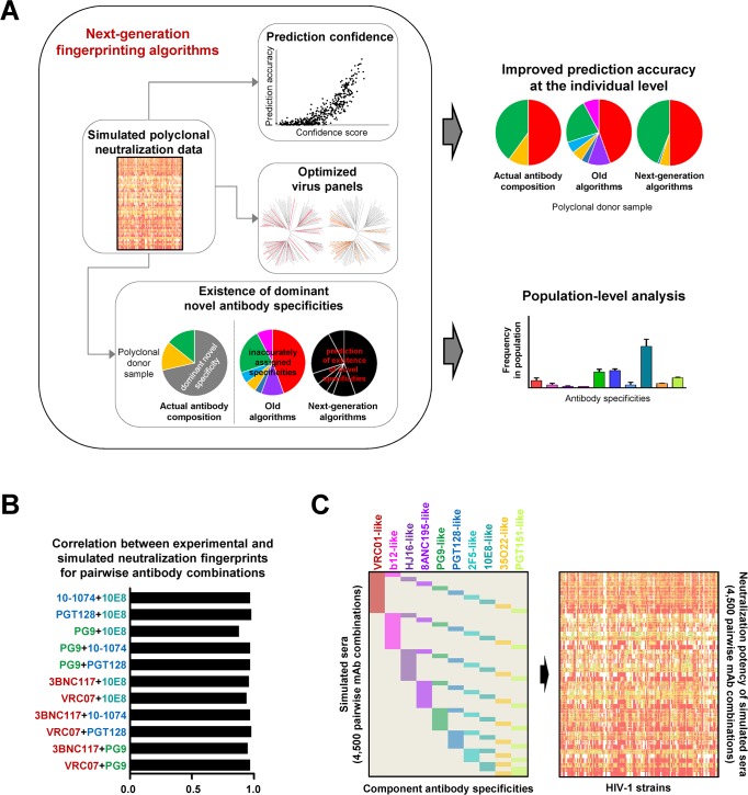
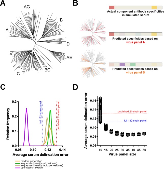
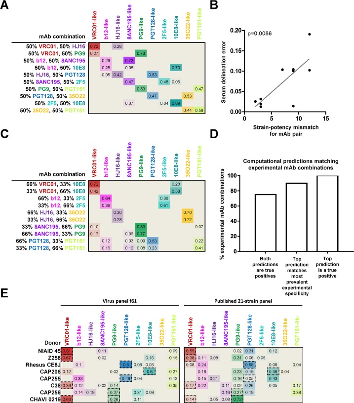
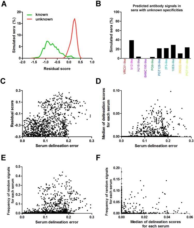
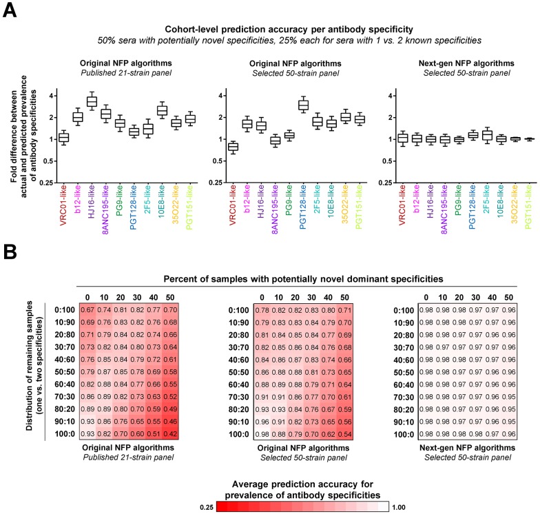
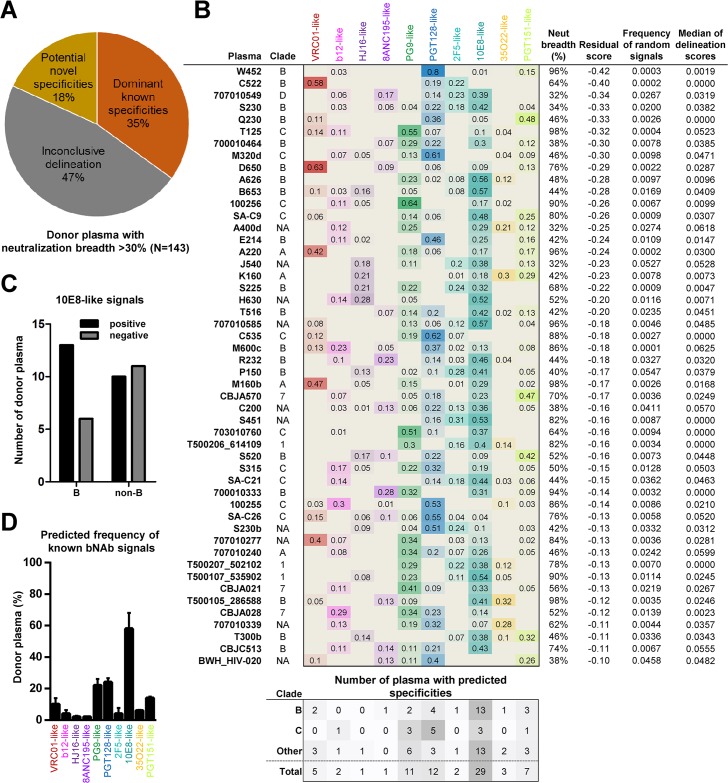
Computational neutralization fingerprinting, NFP, is an efficient and accurate method for predicting the epitope specificities of polyclonal antibody responses to HIV-1 infection. Here, we present next-generation NFP algorithms that substantially improve prediction accuracy for individual donors and enable serologic analysis for entire cohorts. Specifically, we developed algorithms for: (a) selection of optimized virus neutralization panels for NFP analysis, (b) estimation of NFP prediction confidence for each serum sample, and (c) identification of sera with potentially novel epitope specificities. At the individual donor level, the next-generation NFP algorithms particularly improved the ability to detect multiple epitope specificities in a sample, as confirmed both for computationally simulated polyclonal sera and for samples from HIV-infected donors. Specifically, the next-generation NFP algorithms detected multiple specificities in twice as many samples of simulated sera. Further, unlike the first-generation NFP, the new algorithms were able to detect both of the previously confirmed antibody specificities, VRC01-like and PG9-like, in donor CHAVI 0219. At the cohort level, analysis of ~150 broadly neutralizing HIV-infected donor samples suggested a potential connection between clade of infection and types of elicited epitope specificities. Most notably, while 10E8-like antibodies were observed in infections from different clades, an enrichment of such antibodies was predicted for clade B samples. Ultimately, such large-scale analyses of antibody responses to HIV-1 infection can help guide the design of epitope-specific vaccines that are tailored to take into account the prevalence of infecting clades within a specific geographic region. Overall, the next-generation NFP technology will be an important tool for the analysis of broadly neutralizing polyclonal antibody responses against HIV-1.


Conflict of interest statement

The authors have declared that no competing interests exist.

Figures

Fig 1
Fig 1. Next-generation neutralization fingerprinting algorithms for analysis of polyclonal responses against HIV-1.
(A) Schematic of algorithm advances and applications. Large-scale simulations of polyclonal neutralization data were used as a benchmark for the development of the different algorithm components: algorithms for improved confidence in the computational predictions, for selection of optimized virus panels, and for prediction of potentially novel antibody specificities in a serum neutralization signal. Together, these algorithms build the foundation for improving the NFP prediction accuracy at the individual level and enabling NFP analysis at the population (or cohort) level. (B) Spearman correlation coefficients for simulated vs. experimentally determined neutralization data for combinations of antibody pairs. Antibody names are colored according to the respective epitope-specific clusters from (C). (C) Large-scale simulation of polyclonal antibody neutralization, represented as a heatmap based on neutralization potency (white-green-yellow-red).
Fig 2
Fig 2. Identification of improved virus panels for NFP analysis.
(A) Phylogenetic tree of full set of 132 HIV-1 strains used as a source for virus panel selection. (B) Schematic of virus panel evaluation using simulated sera. Heatmaps show the relative prevalence of actual or predicted antibody specificities in a given simulated serum, highlighting the increased accuracy of NFP predictions when using panel A (red strains) vs. panel B (orange strains). (C) Virus panel search methods using four different approaches (colored curves). For each approach, shown are the relative frequencies (y-axis) of observing virus panels of size 20 with different levels of prediction accuracy (x-axis, average serum delineation error, computed over a set of simulated sera for each virus panel). Highlighted are the serum delineation errors for the published 21-strain panel (red) and the full 132-strain panel (blue). (D) Virus panel size vs. prediction accuracy. Each dot represents the average serum delineation error of a single virus panel, with grey bars representing mean and standard deviation for each virus panel size. Shown are the top 5,000 virus panels for a search method and panel size.
Fig 3
Fig 3. Computational comparison of f61, a selected 20-strain virus panel, to the published 21-strain panel.
Both panels were evaluated on the test set of simulated sera. (A) Percent of simulated sera with one or two true positive (red) and one or two+ false positive (blue) signals. (B) Number of simulated sera with true positive (red, out of a maximum of 900) and false positive (blue) signals for each of the ten antibody specificities from the reference set (colored columns).
Fig 4
Fig 4. NFP analysis of mAb combinations and donor sera for virus panel f61.
(A) NFP predictions for ten pairwise mAb combinations (rows), 1:1 ratio. Values shown are the NFP delineation scores for each of ten antibody specificities (columns) in each mAb combination; colored heatmap is proportional to the respective delineation scores. (B) Correlation between serum delineation error and strain-potency mismatch for the ten mAb pairs from (A). (C) NFP predictions for ten pairwise mAb combinations, 2:1 ratio. (D) Agreement between NFP predictions and actual specificities for the mAb combinations in (A,C). (E) NFP analysis of eight donor sera against f61 and the published 21-strain panel. Dotted-line boxes highlight experimentally confirmed specificities.
Fig 5
Fig 5. Prediction of potentially novel specificities and confidence scores in NFP analysis with virus panel f61.
(A) Histograms of residual scores for simulated sera with dominant known (green) vs. unknown (red) specificities. (B) Frequency of predicted known antibody signals (colored) for simulated sera with unknown specificities. (C-E) Relationship between serum delineation error (a measure of NFP prediction accuracy) and three confidence scores: (C) residual scores (for values of at least -0.5), (D) median of the ten delineation scores, and (E) frequency of random signals for a set of simulated sera with dominant known antibody specificities (dots). For each of the three confidence scores, higher values were generally associated with greater serum delineation errors. (F) Complementarity of confidence scores. Shown are the frequency of random signals and median of delineation scores for simulated sera (dots) with residual scores less than -0.1.
Fig 6
Fig 6. Simulation of cohort-level analysis of polyclonal antibody responses.
Three sets of NFP analyses were performed: (left) original NFP algorithms with the published 21-strain panel, (middle) original NFP algorithms with a selected 50-strain panel, and (right) next-generation NFP algorithms with the selected 50-strain panel. (A) Analysis for a simulated sample with 50% sera with potentially novel specificities, and 25% each of sera with 1 or 2 dominant known specificities. For each antibody specificity from the reference set (colored labels), shown are fold differences between actual and predicted prevalence in 1,000 samples of 200 sera each. Boxes represent 25–75 percentiles of the data, with vertical bars corresponding to the 10–90 percentiles, and the median shown as a horizontal line within each box. (B) Analysis for different distributions of simulated sera with known (rows) vs. potentially novel (columns) specificities. Each value shown is the average of the median fold differences for each of the ten specificities given the respective distribution of sera. Values are colored as a red-white heatmap.
Fig 7
Fig 7. Large-scale analysis of HIV-infected donors.
(A) NFP predictions for 143 donor plasma were divided into three groups: plasma with dominant known specificities, with potentially novel specificities, or with inconclusive delineation. (B) Delineated antibody specificities for the 50 plasma with predicted dominant known specificities. Also shown are the clade of infection (A, B, C, D; 1: CRF 01; 7: CRF 07; NA: unknown), neutralization breadth, as well as the three confidence scores, for each sample. The number of positive signals for clades B and C are shown at the bottom, with the ‘Other’ category including both non-B and non-C as well as samples with unknown clades. (C) Prevalence of 10E8-like signals in clade B vs. non-B samples. (D) Overall prevalence of antibody specificities (thick bars) in the 50 samples from (B), and adjusted predictions after the application of the cohort-level NFP algorithm (thin bars). The analysis was based on the selected 50-strain panel from Fig 6.

References

    1. Pancera M, Zhou T, Druz A, Georgiev IS, Soto C, Gorman J, et al. Structure and immune recognition of trimeric pre-fusion HIV-1 Env. Nature. 2014;514(7523):455–61. 10.1038/nature13808 - DOI - PMC - PubMed
    1. Korber B, Gaschen B, Yusim K, Thakallapally R, Kesmir C, Detours V. Evolutionary and immunological implications of contemporary HIV-1 variation. Br Med Bull. 2001;58:19–42. Epub 2001/11/21. - PubMed
    1. Karlsson Hedestam GB, Fouchier RAM, Phogat S, Burton DR, Sodroski J, Wyatt RT. The challenges of eliciting neutralizing antibodies to HIV-1 and to influenza virus. Nat Rev Micro. 2008;6(2):143–55. - PubMed
    1. Georgiev IS, Doria-Rose NA, Zhou T, Kwon YD, Staupe RP, Moquin S, et al. Delineating antibody recognition in polyclonal sera from patterns of HIV-1 isolate neutralization. Science. 2013;340(6133):751–6. Epub 2013/05/11. 10.1126/science.1233989 - DOI - PubMed
    1. Corti D, Langedijk JP, Hinz A, Seaman MS, Vanzetta F, Fernandez-Rodriguez BM, et al. Analysis of memory B cell responses and isolation of novel monoclonal antibodies with neutralizing breadth from HIV-1-infected individuals. PloS one. 2010;5(1):e8805 10.1371/journal.pone.0008805 - DOI - PMC - PubMed

Publication types