

Temporal and spatial regulation of mRNA export: Single particle RNA-imaging provides new tools and insights
- PMID: 28052353
- PMCID: PMC5992323
- DOI: 10.1002/bies.201600124
Temporal and spatial regulation of mRNA export: Single particle RNA-imaging provides new tools and insights
Abstract
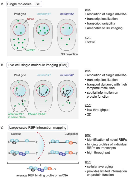
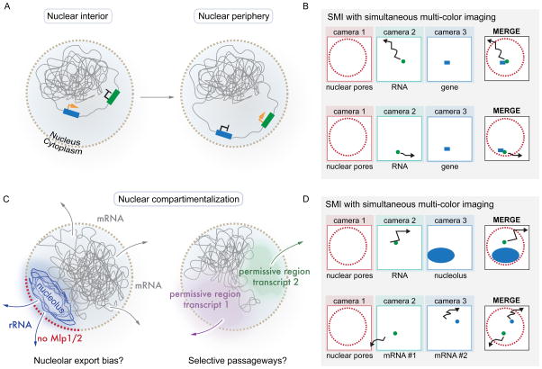
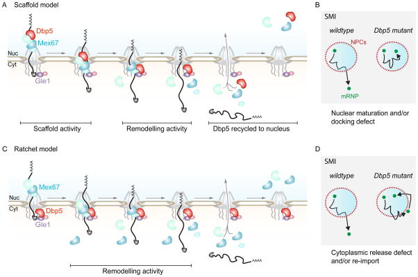
The transport of messenger RNAs (mRNAs) from the nucleus to cytoplasm is an essential step in the gene expression program of all eukaryotes. Recent technological advances in the areas of RNA-labeling, microscopy, and sequencing are leading to novel insights about mRNA biogenesis and export. This includes quantitative single molecule imaging (SMI) of RNA molecules in live cells, which is providing knowledge of the spatial and temporal dynamics of the export process. As this information becomes available, it leads to new questions, the reinterpretation of previous findings, and revised models of mRNA export. In this review, we will briefly highlight some of these recent findings and discuss how live cell SMI approaches may be used to further our current understanding of mRNA export and gene expression.
Keywords: MS2-MCP system; PP7-PCP system; RNA-binding protein; in vivo single molecule imaging; mRNA export; nuclear pore complex.
© 2017 WILEY Periodicals, Inc.
Figures
References
-
- Simon DN, Rout MP. Cancer and the nuclear pore complex. Advances in Exp Med Biol. 2014;773:285–307. - PubMed
Publication types
MeSH terms
Substances
Grants and funding
LinkOut - more resources
Full Text Sources
Other Literature Sources
Research Materials
Miscellaneous
