Clinical Trial
. 2017 Feb 23;61(3):e02197-16.
doi: 10.1128/AAC.02197-16. Print 2017 Mar.

The Oral β-Lactamase SYN-004 (Ribaxamase) Degrades Ceftriaxone Excreted into the Intestine in Phase 2a Clinical Studies

Affiliations
Clinical Trial

The Oral β-Lactamase SYN-004 (Ribaxamase) Degrades Ceftriaxone Excreted into the Intestine in Phase 2a Clinical Studies

John F Kokai-Kun et al. Antimicrob Agents Chemother. .

Abstract

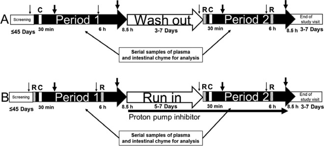
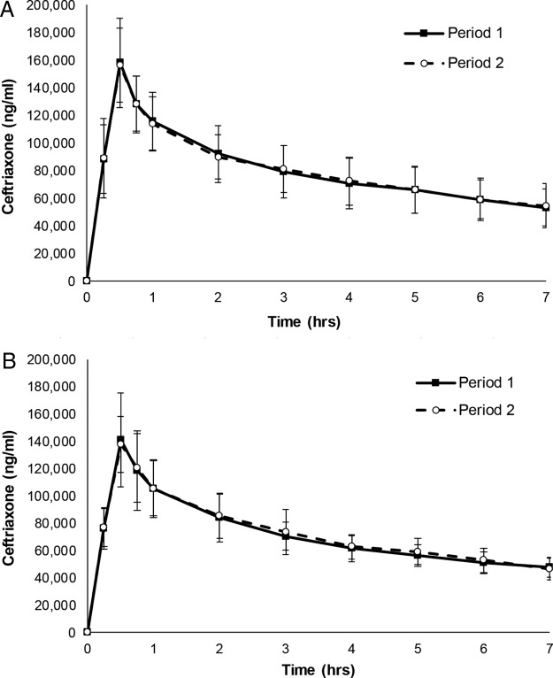
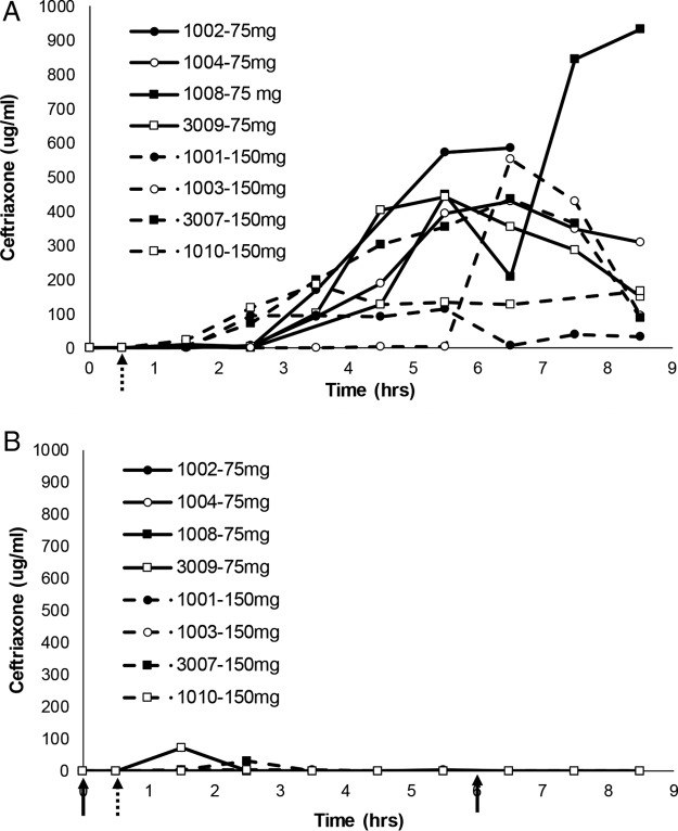
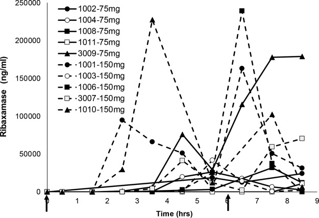
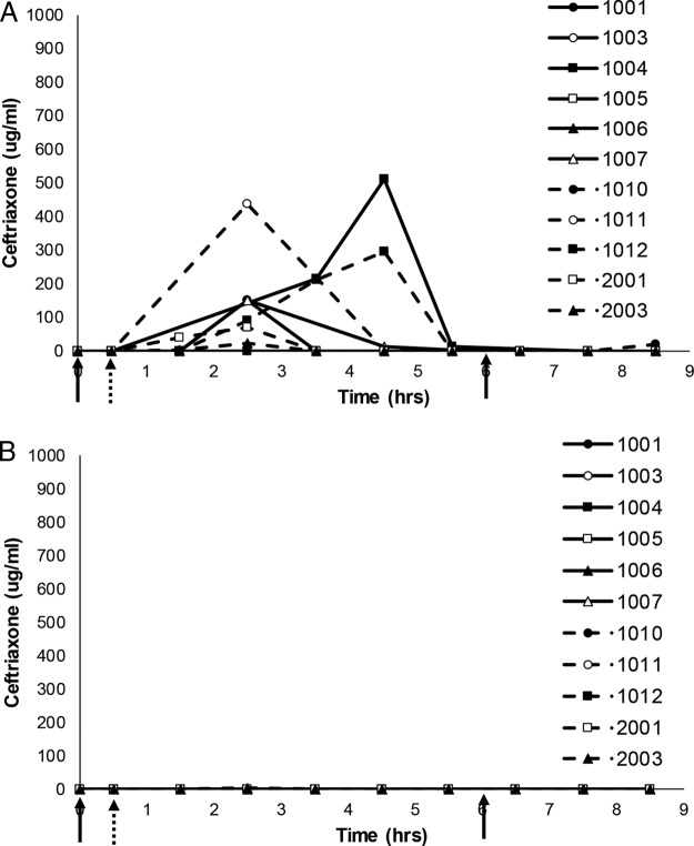
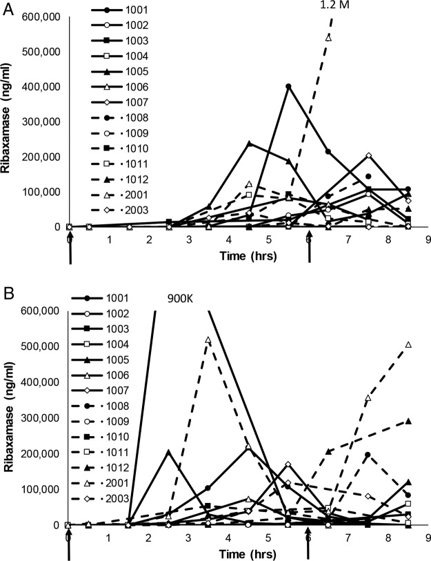
SYN-004 (ribaxamase) is a β-lactamase designed to be orally administered concurrently with intravenous β-lactam antibiotics, including most penicillins and cephalosporins. Ribaxamase's anticipated mechanism of action is to degrade excess β-lactam antibiotic that is excreted into the small intestine. This enzymatic inactivation of excreted antibiotic is expected to protect the gut microbiome from disruption and thus prevent undesirable side effects, including secondary infections such as Clostridium difficile infections, as well as other antibiotic-associated diarrheas. In phase 1 clinical studies, ribaxamase was well tolerated compared to a placebo group and displayed negligible systemic absorption. The two phase 2a clinical studies described here were performed to confirm the mechanism of action of ribaxamase, degradation of β-lactam antibiotics in the human intestine, and were therefore conducted in subjects with functioning ileostomies to allow serial sampling of their intestinal chyme. Ribaxamase fully degraded ceftriaxone to below the level of quantitation in the intestines of all subjects in both studies. Coadministration of oral ribaxamase with intravenous ceftriaxone was also well tolerated, and the plasma pharmacokinetics of ceftriaxone were unchanged by ribaxamase administration. Since ribaxamase is formulated as a pH-dependent, delayed-release formulation, the activity of ribaxamase in the presence of the proton pump inhibitor esomeprazole was examined in the second study; coadministration of these drugs did not adversely affect ribaxamase's ability to degrade ceftriaxone excreted into the intestine. These studies have confirmed the in vivo mechanism of action of ribaxamase, degradation of β-lactam antibiotics in the human intestine (registered at ClinicalTrials.gov under NCT02419001 and NCT02473640).

Keywords: beta-lactamases; ceftriaxone; clinical trials; dysbiosis; gut microbiome; oral administration; protection.


Figures

FIG 1
FIG 1
Study design for the two clinical studies conducted in subjects with ileostomies. (A) Study 1; (B) study 2. Patients were screened for the studies up to 45 days prior to period 1. An intravenous infusion (30-min infusion) of ceftriaxone (1 g) was administered at 30 min of each period as indicated (C), and oral ribaxamase (75 or 150 mg in study 1 and 150 mg in study 2) was administered at 0 min and 6 h as indicated (R). Small nonfatty meals were provided at 1 h prior to and 5 h after the infusion (small arrows) and full meals at 2 and 7.5 h postinfusion (thick arrows). Subjects were required to drink water or apple juice during periods 1 and 2 to promote intestinal chyme production. A washout period of 3 to 7 days separated period 1 and period 2 of study 1, and in study 2, a run-in period of 5 to 7 days occurred during which subjects self-administered 40 mg of oral esomeprazole daily in the morning. In study 2, subjects also received 40 mg of esomeprazole 1 h prior to the first dose of ribaxamase in period 2. In both studies, serial plasma and chyme samples were collected for analysis during periods 1 and 2, and an end-of-study visit occurred 3 to 7 days after period 2.
FIG 2
FIG 2
Comparison of ceftriaxone concentrations in plasma in periods 1 and 2 in the two studies. The graphs display the mean plasma ceftriaxone concentration (± the standard deviations [SD]) over time for 10 subjects from study 1 (A) and 14 subjects from study 2 (B). Study 1 period 1, i.v. ceftriaxone only; period 2, i.v. ceftriaxone + oral ribaxamase. Study 2 period 1, i.v. ceftriaxone + oral ribaxamase; period 2, i.v. ceftriaxone + oral ribaxamase + esomeprazole. The 0-h time points on the graphs represent the ceftriaxone i.v. infusion start.
FIG 3
FIG 3
Comparison of ceftriaxone concentrations in intestinal chyme for eight subjects from study 1. Serial chyme samples (as available) were collected and analyzed for their ceftriaxone concentrations over time in periods 1 (A) and 2 (B) of study 1. The data show individual ceftriaxone concentration curves (assay lower limit of quantitation, 1 μg/ml) for eight subjects (subject numbers indicated on the figure) over time. The 30-min i.v. ceftriaxone (1 g) infusion began at the hatched arrow, while 75 or 150 mg (as randomized and indicated on the figures) of oral ribaxamase was administered at the solid arrows in period 2. Missing data points indicate that no chyme was available for collection at that time point. Two subjects from this study were excluded from this figure (see the text and Fig. S1 in the supplemental material).
FIG 4
FIG 4
Ribaxamase concentrations in intestinal chyme for ten subjects from period 2 of study 1. Serial chyme samples (as available) were collected and analyzed for their ribaxamase concentrations over time in period 2 of study 1. The data show individual concentration curves (assay LLOQ, 10 ng/ml) for 10 subjects (subject numbers are indicated in the figure) over time. Two doses of oral ribaxamase were administered (75 or 150 mg, as randomized and indicated on the figure) at the solid arrows. Missing data points indicate that no chyme was available for collection at that time point.
FIG 5
FIG 5
Comparison of ceftriaxone concentrations in intestinal chyme for eleven subjects from study 2. Serial chyme samples (as available) were collected and analyzed for their ceftriaxone concentrations over time in periods 1 (A) and 2 (B) of study 2. The data show individual ceftriaxone concentration curves (assay LLOQ, 1 μg/ml) for 11 subjects (subject numbers are indicated in the figure) over time. The 30-min ceftriaxone (1 g) infusion began at the hatched arrow, while 150 mg of oral ribaxamase was administered at the solid arrows. Period 2 was when esomeprazole was present. Missing data points indicate that no chyme was available for collection at that time point. Three subjects from this study were excluded from this figure (see the text and Fig. S2 in the supplemental material).
FIG 6
FIG 6
Ribaxamase concentrations in intestinal chyme for fourteen subjects from study 2. Serial chyme samples (as available) were collected and analyzed for their ribaxamase concentrations over time in hours in periods 1 (A) and 2 (B) of study 2. The data show individual concentration curves (assay LLOQ, 10 ng/ml) for 14 subjects (subject numbers are indicated in the figure) over time. Two doses of oral ribaxamase were administered (150 mg/dose) at the solid arrows. Missing data indicate that no chyme was available for collection at that time point. Period 2 is when esomeprazole was present. Both graphs were truncated for clarity, and the numbers indicate the peak concentrations for the two subjects with truncated profiles.

References

    1. Kinross JM, Darzi AW, Nicholson JK. 2011. Gut microbiome-host interactions in health and disease. Genome Med 3:14. doi:10.1186/gm228. - DOI - PMC - PubMed
    1. Konkel L, Danska J, Mazmanian S, Chadwick L. 2013. The environment within: exploring the role of the gut microbiome in health and disease. Environ Health Perspect 121:a276–a281. doi:10.1289/ehp.121-A276. - DOI - PMC - PubMed
    1. Britton RA, Young VB. 2014. Role of the intestinal microbiota in resistance to colonization by Clostridium difficile. Gastroenterology 146:1547–1553. doi:10.1053/j.gastro.2014.01.059. - DOI - PMC - PubMed
    1. Chalmers JD, Akram AR, Singanayagam A, Wilcox MH, Hill AT. 2016. Risk factors for Clostridium difficile infection in hospitalized patients with community-acquired pneumonia. J Infect 73:45–53. doi:10.1016/j.jinf.2016.04.008. - DOI - PubMed
    1. Crowther GS, Wilcox MH. 2015. Antibiotic therapy and Clostridium difficile infection—primum non nocere—first do no harm. Infect Drug Resist 8:333–337. doi:10.2147/IDR.S87224. - DOI - PMC - PubMed

Publication types

MeSH terms

Associated data