. 2017 Feb 28;91(6):e02220-16.
doi: 10.1128/JVI.02220-16. Print 2017 Mar 15.

Heat Shock Protein 90 Ensures Efficient Mumps Virus Replication by Assisting with Viral Polymerase Complex Formation

Affiliations

Heat Shock Protein 90 Ensures Efficient Mumps Virus Replication by Assisting with Viral Polymerase Complex Formation

Hiroshi Katoh et al. J Virol. .

Abstract

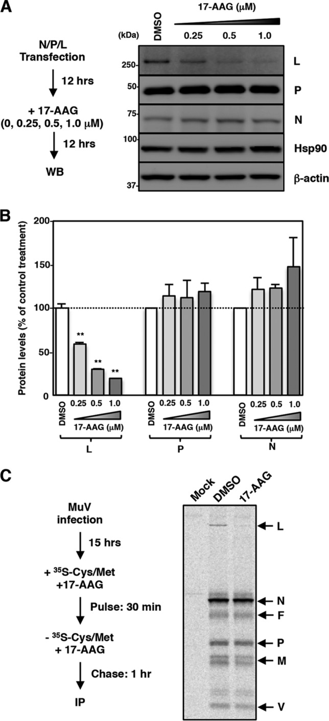
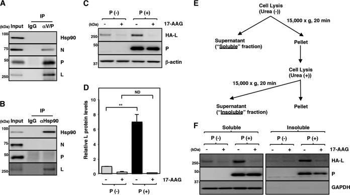
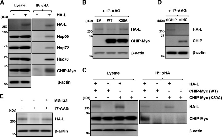
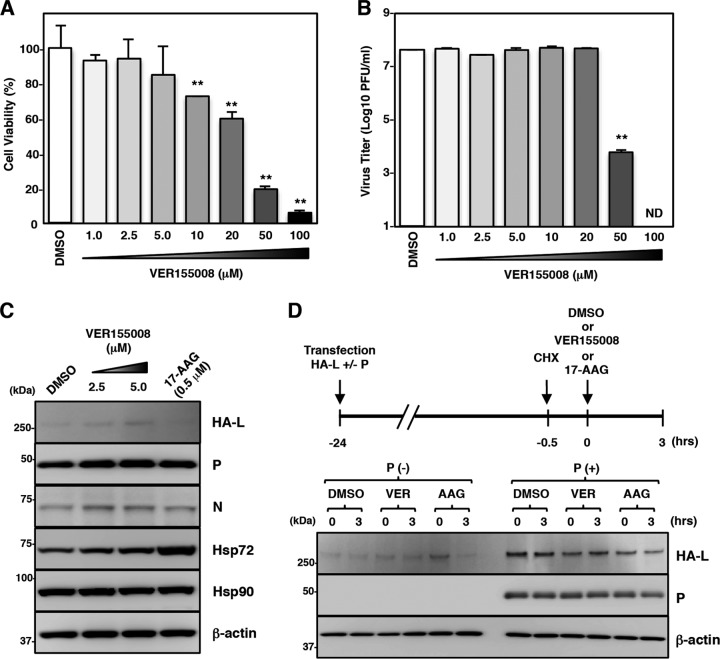
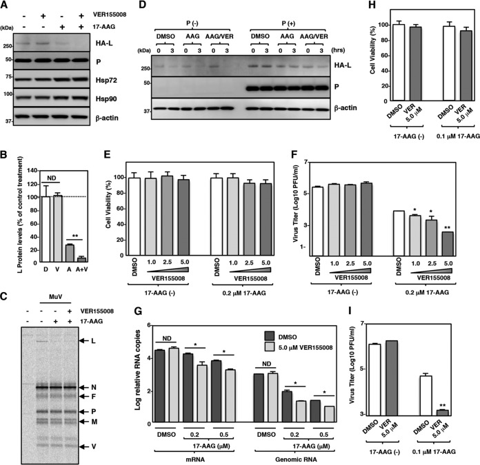
Paramyxoviral RNAs are synthesized by a viral RNA-dependent RNA polymerase (RdRp) consisting of the large (L) protein and its cofactor phosphoprotein (P protein). The L protein is a multifunctional protein that catalyzes RNA synthesis, mRNA capping, and mRNA polyadenylation. Growing evidence shows that the stability of several paramyxovirus L proteins is regulated by heat shock protein 90 (Hsp90). In this study, we demonstrated that Hsp90 activity was important for mumps virus (MuV) replication. The Hsp90 activity was required for L-protein stability and activity because an Hsp90-specific inhibitor, 17-allylamino-17-demethoxygeldanamycin (17-AAG), destabilized the MuV L protein and suppressed viral RNA synthesis. However, once the L protein formed a mature polymerase complex with the P protein, Hsp90 activity was no longer required for the stability and activity of the L protein. When the Hsp90 activity was inhibited, the MuV L protein was degraded through the CHIP (C terminus of Hsp70-interacting protein)-mediated proteasomal pathway. High concentrations of 17-AAG showed strong cytotoxicity to certain cell types, but combined use of an Hsp70 inhibitor, VER155008, potentiated degradation of the L protein, allowing a sufficient reduction of 17-AAG concentration to block MuV replication with minimum cytotoxicity. Regulation of the L protein by Hsp90 and Hsp70 chaperones was also demonstrated for another paramyxovirus, the measles virus. Collectively, our data show that the Hsp90/Hsp70 chaperone machinery assists in the maturation of the paramyxovirus L protein and thereby in the formation of a mature RdRp complex and efficient viral replication.IMPORTANCE Heat shock protein 90 (Hsp90) is nearly universally required for viral protein homeostasis. Here, we report that Hsp90 activity is required for efficient propagation of mumps virus (MuV). Hsp90 functions in the maintenance of the catalytic subunit of viral polymerase, the large (L) protein, prior to formation of a mature polymerase complex with the polymerase cofactor of L, phosphoprotein. Hsp70 collaborates with Hsp90 to regulate biogenesis of the MuV L protein. The functions of these chaperones on the viral polymerase may be common among paramyxoviruses because the L protein of measles virus is also similarly regulated. Our data provide important insights into the molecular mechanisms of paramyxovirus polymerase maturation as well as a basis for the development of novel antiviral drugs.

Keywords: heat shock protein 90; large protein; mumps virus; polymerase.


Figures

FIG 1
FIG 1
Hsp90 activity is required for MuV propagation. (A) Schematic of the rOdate and rOdate/AcGFP genes. SH, small hydrophobic gene. (B) Vero cells infected with rOdate or rOdate/AcGFP were observed under phase-contrast and a fluorescence microscopes at 48 hpi. (C) Vero cells were infected with rOdate or rOdate/AcGFP at an MOI of 0.01. The supernatants were collected at 24, 48, 72, and 96 hpi, and the infectious titers were determined by plaque assay. (D) Vero cells were treated with the indicated concentrations of 17-AAG for 96 h, and then cell viability was determined and calculated as a percentage of the viability of cells treated with DMSO. (E and F) Vero cells were infected with rOdate/AcGFP at an MOI of 0.01 and treated with the indicated concentrations of 17-AAG. At 96 hpi, the cells were observed under a fluorescence microscope (E), and the infectious titers in the supernatants were determined (F). (G) A549 cells were treated with 0.1 and 0.2 μM 17-AAG for 96 h, and then cell viability was determined and calculated as a percentage of the viability of cells treated with DMSO. (H and I) A549 cells were infected with rOdate/AcGFP at an MOI of 0.01 and treated with 0.1 μM 17-AAG. At 96 hpi, the cells were observed under a fluorescence microscope (H), and the infectious titers in the supernatants were determined (I). Error bars indicate the standard deviations of triplicates. The significance of differences between the means was determined by Student's t test. *, P < 0.05; **, P < 0.01. ND, not detected; DAPI, 4′,6′-diamidino-2-phenylindole.
FIG 2
FIG 2
Hsp90 activity is required for MuV RNA synthesis after the secondary transcription phase. (A) Schematic of the experimental procedures showing the times of RNA collection and 17-AAG treatment after virus infection. (B and C) Vero cells were infected with MuV at an MOI of 5.0. At the indicated times postinfection, total cellular RNA was extracted and subjected to an RT reaction using oligo(dT) and strand-specific primers. The levels of N mRNA and genomic RNA were determined by qPCR. The values were normalized to the level of the control gene HPRT1 and are shown as the log fold change. Three independent experiments were performed, and the most representative data are shown. (D and E) Vero cells were infected with MuV at an MOI of 5.0 and cultured with or without 50 μg/ml of CHX and 0.5 μM 17-AAG or DMSO. At 0 and 24 hpi, total cellular RNA was extracted and subjected to an RT reaction using oligo(dT) and strand-specific primers. The levels of N mRNA and genomic RNA were determined by qPCR. The values were normalized to the value for the control gene HPRT1. (F) BHK/T7-9 cells were treated with the indicated concentrations of 17-AAG for 48 h, and then cell viability was determined and calculated as a percentage of the viability of cells treated with DMSO. (G) BHK/T7-9 cells were transfected with the minigenome plasmid (pFL-MuV-MG) and three support plasmids (pCR-N, -P, and -L). At 4 h posttransfection, the medium was replaced with fresh medium containing 17-AAG, as indicated in the figure. pCR-L was omitted from the transfection mixture to establish a negative control. The luciferase activity in DMSO-treated cells was set to 100%. Error bars indicate the standard deviations of triplicates. The significance of differences between the means was determined by Student's t test. *, P < 0.05; **, P < 0.01. ND, no significant difference.
FIG 3
FIG 3
MuV L protein is a client of Hsp90. (A and B) 293T cells were cotransfected with three plasmids, pCAGGS-N, -P, and -L, and the medium was replaced with fresh medium containing the indicated concentrations of 17-AAG at 12 h posttransfection. At 24 h posttransfection, the cell lysates were subjected to immunoblotting with the indicated antibodies (A). The relative band intensities of each viral protein were normalized by the β-actin level, and the relative expression levels of each viral protein based on the levels of DMSO-treated cells are shown in panel B. (C) Vero cells were infected with MuV at an MOI of 5.0. At 15 hpi, the cells were radiolabeled for 30 min with [35S]methionine-cysteine and incubated in nonradioactive medium for 1 h before lysis and analysis by immunoprecipitation with an anti-MuV antibody. 17-AAG was incubated at 0.5 μM during the pulse and chase periods. WB, Western blotting; IP, immunoprecipitation.
FIG 4
FIG 4
The maturation of the L protein is sequentially regulated by Hsp90 and the P protein. (A and B) At 24 hpi, Vero cells infected with MuV at an MOI of 5.0 were subjected to immunoprecipitation with anti-MuV V/P or anti-Hsp90 antibody and immunoblotting with the indicated antibodies. (C and D) 293T cells were transfected with pCAGGS-HA-MuV-L alone or cotransfected with pCAGGS-HA-MuV-L and pCAGGS-P, and the medium was replaced with fresh medium containing 0.5 μM 17-AAG at 12 h posttransfection. At 24 h posttransfection, the cell lysates were prepared using a urea-free cell lysis buffer and subjected to immunoblotting with the indicated antibodies. The relative band intensities of L protein were normalized to the β-actin level, and the relative expression levels of L protein based on the levels of DMSO-treated cells without P are shown (D). (E and F) At 24 h posttransfection, soluble and insoluble fractions of the cells were prepared as indicated in panel E and subjected to immunoblotting with the antibodies indicated in panel F. Error bars indicate the standard deviations of triplicates. The significance of differences between the means was determined by Student's t test. **, P < 0.01. ND, no significant difference.
FIG 5
FIG 5
Hsp90 is dispensable for stability of the L protein and the polymerase activity after an interaction with the P protein. (A and B) 293T cells were transfected with pCAGGS-HA-MuV-L alone or cotransfected with pCAGGS-HA-MuV-L and pCAGGS-P. At 23.5 h posttransfection, the cells were treated with 50 μg/ml CHX for 30 min. Then, 0.5 μM 17-AAG was added to the cells, and the cell lysates were collected at the indicated times and subjected to immunoblotting. The relative band intensities of L and P proteins were normalized to the β-actin level, and the relative expression levels of each viral protein based on the levels of cells collected at 0 h are shown in panel B. (C) Vero cells were infected with MuV at an MOI of 5.0. At 11.5 hpi, the cells were treated with 50 μg/ml of CHX for 30 min. Then, 0.5 μM 17-AAG was added to the cells, and total cellular RNA was extracted at the indicated times and subjected to an RT reaction using oligo(dT) and strand-specific primers. The levels of N mRNA and genomic RNA were determined by qPCR, and the values were normalized to the level of the control gene HPRT1 and shown as log fold changes. Error bars indicate the standard deviations of triplicates. The significance of differences between the means was determined by Student's t test. **, P < 0.01. ND, no significant difference.
FIG 6
FIG 6
Disruption of Hsp90 function induces degradation of the MuV L protein through a CHIP-mediated proteasomal pathway. (A) 293T cells were transfected with pcDNA-CHIP-Myc/His (WT) and/or pCAGGS-HA-MuV-L. At 24 h posttransfection, the cell lysates were subjected to immunoprecipitation with anti-HA antibody and to immunoblotting with the indicated antibodies. (B) 293T cells were cotransfected with pCAGGS-HA-MuV-L and pcDNA-CHIP-Myc/His (WT or K30A). At 24 h posttransfection, the cells were treated with 0.5 μM 17-AAG for 4 h, and then the cell lysates were subjected to immunoblotting with the indicated antibodies. EV, empty vector. (C) HA-MuV-L was coexpressed with CHIP-Myc (WT or K30A) in 293T cells, immunoprecipitated with anti-HA antibody, and immunoblotted with the indicated antibodies. (D) At 48 h posttransfection with either siCHIP or siNC, 293T cells were transfected with pCAGGS-HA-MuV-L. After 24 h, the cells were treated with 0.5 μM 17-AAG for 4 h, and then the cell lysates were subjected to immunoblotting with the indicated antibodies. (E) 293T cells were cotransfected with pCAGGS-HA-MuV-L and pcDNA-CHIP-Myc/His (WT). At 24 h posttransfection, the cells were treated with 0.5 μM 17-AAG and/or 10 μM MG-132 for 4 h, and then the cell lysates were subjected to immunoblotting with anti-HA and anti-β-actin antibodies.
FIG 7
FIG 7
An Hsp70 inhibitor does not affect MuV propagation or the L protein expression. (A) Vero cells were treated with the indicated concentrations of VER155008 for 96 h, and then cell viability was determined and calculated as a percentage of the viability of cells treated with DMSO. (B) Vero cells were infected with rOdate/AcGFP at an MOI of 0.01 and treated with the indicated concentrations of VER155008. At 96 hpi, the infectious titers in the supernatants were determined. (C) 293T cells were cotransfected with three plasmids, pCAGGS-N, -P, and -L, and the medium was replaced with fresh medium containing 2.5 or 5.0 μM VER155008 at 12 h posttransfection. At 24 h posttransfection, the cell lysates were subjected to immunoblotting with the indicated antibodies. (D) 293T cells were transfected with pCAGGS-HA-MuV-L alone or cotransfected with pCAGGS-HA-MuV-L and pCAGGS-P. At 23.5 h posttransfection, the cells were treated with 50 μg/ml CHX for 30 min. Then, 5.0 μM VER155008 (VER) or 0.5 μM 17-AAG (AAG) was added to the cells for 3 h, and the cell lysates were collected and subjected to immunoblotting. Error bars indicate the standard deviations of triplicates. The significance of differences between the means was determined by Student's t test. **, P < 0.01. ND, not detected.
FIG 8
FIG 8
The Hsp70 inhibitor potentiates degradation of the MuV L protein under Hsp90 inhibitor treatment. (A and B) 293T cells were cotransfected with pCAGGS-HA-MuV-L and pCAGGS-P, and the medium was replaced with fresh medium containing 0.5 μM 17-AAG and/or 5.0 μM VER155008 at 12 h posttransfection. At 24 h posttransfection, the cell lysates were subjected to immunoblotting (A) with the indicated antibodies. The relative band intensities of HA-L protein were normalized to the β-actin level, and the relative expression levels of L protein based on the levels of DMSO-treated cells are shown in panel B. D, DMSO; V, VER155008; A, 17-AAG; V+A, VER155008 and 17-AAG. (C) Vero cells were infected with MuV at an MOI of 5.0. At 15 hpi, the cells were radiolabeled for 30 min with [35S]methionine-cysteine and incubated in nonradioactive medium for 1 h before lysis and analysis by immunoprecipitation with an anti-MuV antibody. 17-AAG (0.5 μM) and/or VER155008 (5.0 μM) was incubated during the pulse and chase periods. (D) 293T cells were transfected with pCAGGS-HA-MuV-L alone or cotransfected with pCAGGS-HA-MuV-L and pCAGGS-P. At 23.5 h posttransfection, the cells were treated with 50 μg/ml of CHX for 30 min. Then, 17-AAG (0.5 μM) and/or VER155008 (5.0 μM) was added to the cells for 3 h, and the cell lysates were collected and subjected to immunoblotting. AAG,17-AAG; AAG/VER, 17-AAG and VER155008. (E) Vero cells were treated with the indicated concentrations of VER155008 with/without 0.2 μM 17-AAG for 96 h, and then cell viability was determined and calculated as a percentage of the viability of cells treated with DMSO alone. (F) Vero cells were infected with rOdate/AcGFP at an MOI of 0.01 and treated with the indicated concentrations of VER155008 with/without 0.2 μM 17-AAG. At 96 hpi, the infectious titers in the supernatants were determined. (G) Vero cells were infected with MuV at an MOI of 5.0 and treated with 5.0 μM VER155008 and the indicated concentrations of 17-AAG. At 24 hpi, total cellular RNA was extracted and subjected to an RT reaction using oligo(dT) and strand-specific primers. The levels of N mRNA and genomic RNA were determined by qPCR, and the values were normalized to the value for the control gene HPRT1 and are shown as log fold changes. (H) A549 cells were treated with 5.0 μM VER155008 with/without 0.1 μM 17-AAG for 96 h, and then cell viability was determined and calculated as a percentage of the viability of cells treated with DMSO alone. (I) A549 cells were infected with rOdate/AcGFP at an MOI of 0.01 and treated with 5.0 μM VER155008 with/without 0.1 μM 17-AAG. At 96 hpi, the infectious titers in the supernatants were determined. Error bars indicate the standard deviations of triplicates. The significance of differences between the means was determined by Student's t test. *, P < 0.05; **, P < 0.01. ND, no significant difference.
FIG 9
FIG 9
Hsp70 and CHIP are also associated with measles virus (MeV) L protein and involved in MeV infection. (A) 293T cells were transfected with pCAGGS-HA-MeV-L, and the medium was replaced with fresh medium containing 0.5 μM 17-AAG at 12 h posttransfection. At 24 h posttransfection, the cell lysates were subjected to immunoblotting with anti-HA and anti-β-actin antibodies. (B) 293T cells were transfected with pcDNA-CHIP-Myc/His (WT) and/or pCAGGS-HA-MeV-L. At 24 h posttransfection, the cell lysates were subjected to immunoprecipitation with anti-HA antibody and to immunoblotting with the indicated antibodies. (C) Vero/hSLAM cells were infected with IC323-EGFP at an MOI of 0.01 and treated with the indicated concentrations of VER155008 with/without 0.2 μM 17-AAG. At 72 hpi, the infectious titers in the supernatants were determined. Error bars indicate the standard deviations of triplicates. ND, not detected.

Similar articles

Cited by

References

    1. Hviid A, Rubin S, Muhlemann K. 2008. Mumps. Lancet 371:932–944. doi: 10.1016/S0140-6736(08)60419-5. - DOI - PubMed
    1. Lamb RA, Parks GD. 2006. Paramyxoviridae: the viruses and their replication, p 1449–1496. In Knipe DM, Howley PM, Griffin DE, Lamb RA, Martin MA, Roizman B, Straus SE (ed), Fields virology, 5th ed Lippincott Williams & Wilkins, Philadelphia, PA.
    1. Whelan SP, Barr JN, Wertz GW. 2004. Transcription and replication of nonsegmented negative-strand RNA viruses. Curr Top Microbiol Immunol 283:61–119. - PubMed
    1. Emerson SU, Wagner RR. 1973. L protein requirement for in vitro RNA synthesis by vesicular stomatitis virus. J Virol 12:1325–1335. - PMC - PubMed
    1. Hercyk N, Horikami SM, Moyer SA. 1988. The vesicular stomatitis virus L protein possesses the mRNA methyltransferase activities. Virology 163:222–225. doi: 10.1016/0042-6822(88)90253-X. - DOI - PubMed

Substances

LinkOut - more resources