. 2017 Feb 28;91(6):e02322-16.
doi: 10.1128/JVI.02322-16. Print 2017 Mar 15.

5'-Phospho-RNA Acceptor Specificity of GDP Polyribonucleotidyltransferase of Vesicular Stomatitis Virus in mRNA Capping

Affiliations

5'-Phospho-RNA Acceptor Specificity of GDP Polyribonucleotidyltransferase of Vesicular Stomatitis Virus in mRNA Capping

Minako Ogino et al. J Virol. .

Abstract

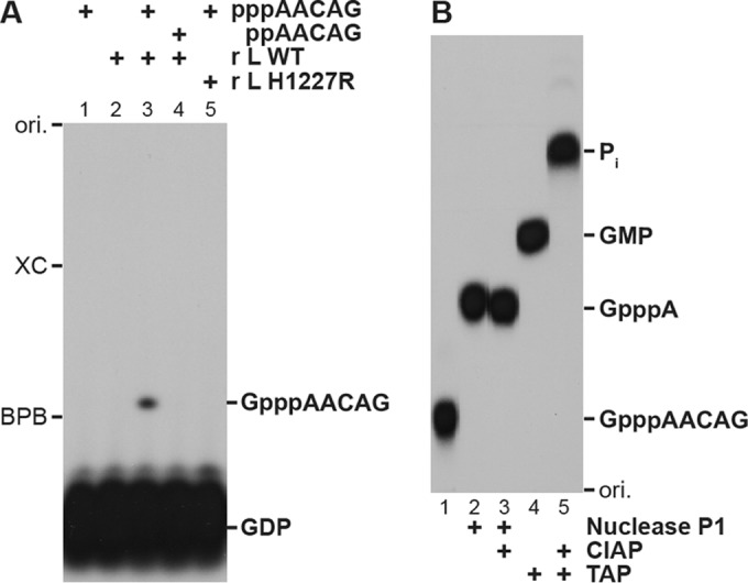
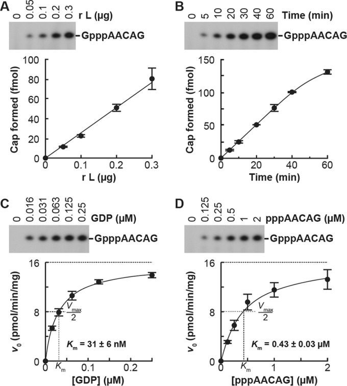
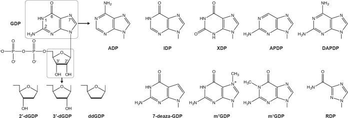
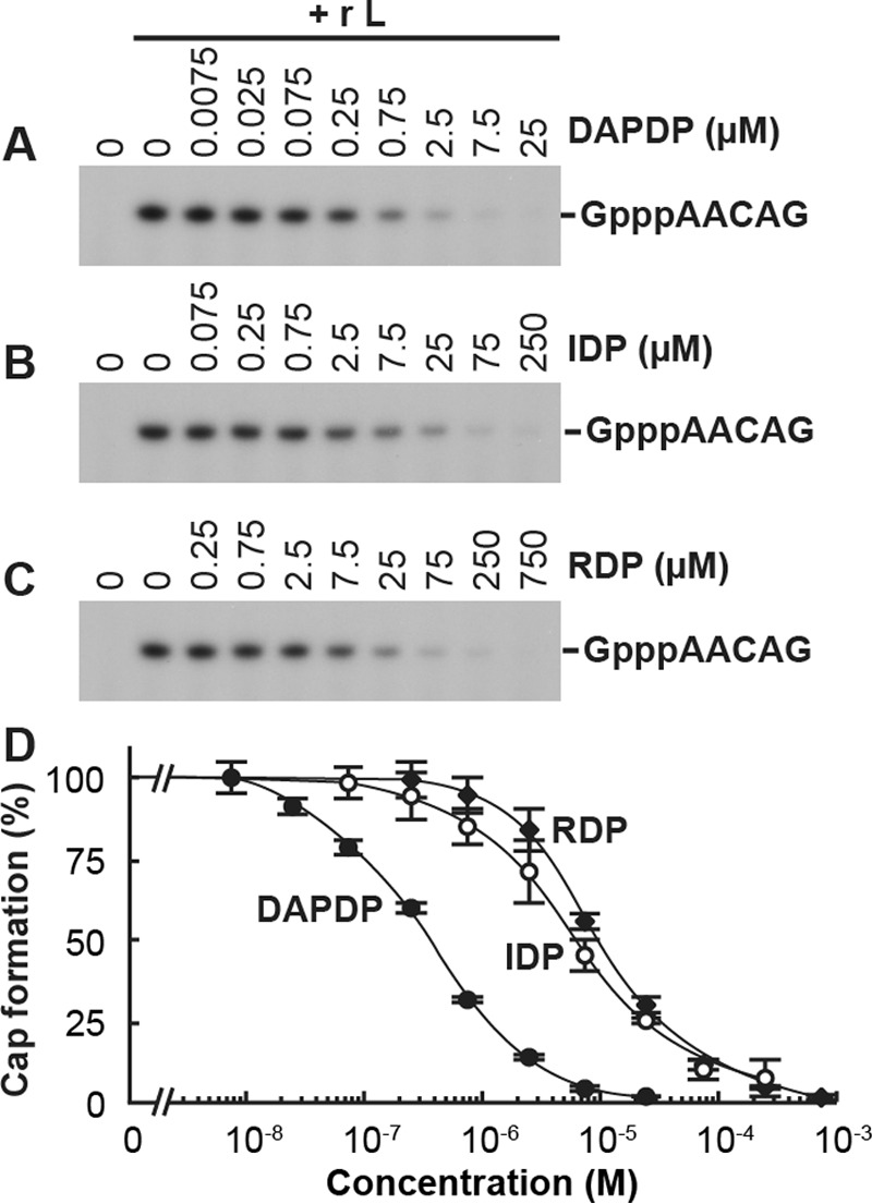
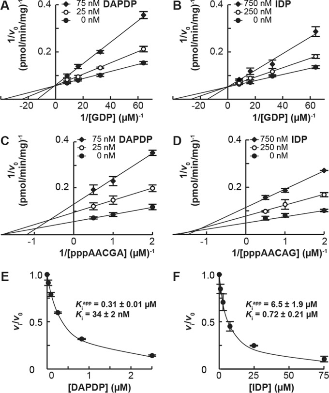
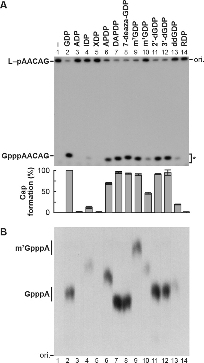
The GDP polyribonucleotidyltransferase (PRNTase) domain of the multifunctional L protein of rhabdoviruses, such as vesicular stomatitis virus (VSV) and rabies virus, catalyzes the transfer of 5'-phospho-RNA (pRNA) from 5'-triphospho-RNA (pppRNA) to GDP via a covalent enzyme-pRNA intermediate to generate a 5'-cap structure (GpppA). Here, using an improved oligo-RNA capping assay with the VSV L protein, we showed that the Michaelis constants for GDP and pppAACAG (VSV mRNA-start sequence) are 0.03 and 0.4 μM, respectively. A competition assay between GDP and GDP analogues in the GpppA formation and pRNA transfer assay using GDP analogues as pRNA acceptors indicated that the PRNTase domain recognizes the C-2-amino group, but not the C-6-oxo group, N-1-hydrogen, or N-7-nitrogen, of GDP for the cap formation. 2,6-Diaminopurine-riboside (DAP), 7-deazaguanosine (7-deaza-G), and 7-methylguanosine (m7G) diphosphates efficiently accepted pRNA, resulting in the formation of DAPpppA, 7-deaza-GpppA, and m7GpppA (cap 0), respectively. Furthermore, either the 2'- or 3'-hydroxyl group of GDP was found to be required for efficient pRNA transfer. A 5'-diphosphate form of antiviral ribavirin weakly inhibited the GpppA formation but did not act as a pRNA acceptor. These results indicate that the PRNTase domain has a unique guanosine-binding mode different from that of eukaryotic mRNA capping enzyme, guanylyltransferase. IMPORTANCE mRNAs of nonsegmented negative-strand (NNS) RNA viruses, such as VSV, possess a fully methylated cap structure, which is required for mRNA stability, efficient translation, and evasion of antiviral innate immunity in host cells. GDP polyribonucleotidyltransferase (PRNTase) is an unconventional mRNA capping enzyme of NNS RNA viruses that is distinct from the eukaryotic mRNA capping enzyme, guanylyltransferase. In this study, we studied the pRNA acceptor specificity of VSV PRNTase using various GDP analogues and identified chemical groups of GDP as essential for the substrate activity. The findings presented here are useful not only for understanding the mechanism of the substrate recognition with PRNTase but also for designing antiviral agents targeting this enzyme.

Keywords: GDP polyribonucleotidyltransferase; L protein; cap structure; mRNA capping; nonsegmented negative-strand RNA viruses; rabies virus; vesicular stomatitis virus.


Figures

FIG 1
FIG 1
Improved oligo-RNA capping assay with the recombinant VSV L protein. (A) [α-32P]GDP (0.25 μM) was incubated in capping reaction mixtures with the indicated RNA (2.5 μM) and/or recombinant VSV L protein (0.2 μg, wild type [WT] or cap-defective mutant [H1227R]) for 20 min. The reaction mixtures were directly analyzed by 20% urea-PAGE followed by autoradiography. (B) [32P]GpppAACAG synthesized by the WT L protein was purified from a polyacrylamide gel and digested with nuclease P1, calf intestine alkaline phosphatase (CIAP), and/or tobacco acid pyrophosphatase (TAP). The digests were analyzed by PEI-cellulose TLC followed by autoradiography. ori., origin.
FIG 2
FIG 2
Kinetic characteristics of the GpppA formation with the VSV L protein. (A) Various amounts of the recombinant VSV L protein were incubated with 0.25 μM [α-32P]GDP and 2.5 μM pppAACGA for 20 min. The resulting GpppAACAG was analyzed as described for Fig. 1A (upper panel). The autoradiogram is a representative of three independent experiments. Amounts of GpppA formed on the RNA were determined by counting the 32P radioactivities of the indicated bands and plotted against the protein amounts (lower panel). Symbols and error bars represent the means and standard deviations, respectively. (B) The time course of the GpppA formation was determined with 0.25 μM [α-32P]GDP, 2.5 μM pppAACGA, and 0.2 μg of the recombinant VSV L protein. (C and D) The recombinant VSV L protein (0.2 μg) was incubated with various concentrations of [α-32P]GDP and 2.5 μM pppAACGA (C) or 0.25 μM [α-32P]GDP and various concentrations of pppAACGA (D) for 20 min. The initial velocity (v0) of the GpppA formation (pmol/min/mg protein) was plotted against the substrate concentrations. The curved lines represent the best fits to the Michaelis-Menten equation. Km values for respective substrates were estimated as described in Materials and Methods.
FIG 3
FIG 3
Structures of GDP analogues. The structure of GDP is shown with numbered positions of some base and ribose ring atoms. Nucleoside 5′-diphosphates with the indicated base or ribose were used in this study. Their full names and abbreviations are listed in Table 1.
FIG 4
FIG 4
GDP analogues inhibit the GpppA formation catalyzed by the VSV L protein. The oligo-RNA capping assay was performed with 0.25 μM [α-32P]GDP and 2.5 μM pppAACGA in the presence or absence of various concentrations of 2,6-diaminopurine-riboside 5′-diphosphate (DAPDP) (A), IDP (B), or ribavirin 5′-diphosphate (RDP) (C). The resulting GpppAACAG was analyzed as described for Fig. 1A. The results are expressed as percentages of the GpppA formation activity observed in the absence of the inhibitors (set to 100%) (D). Symbols and error bars represent the means and standard deviations, respectively, for three independent experiments.
FIG 5
FIG 5
Competitive inhibition of the GpppA formation against GDP by DAPDP and IDP. (A and B) The oligo-RNA capping assay was performed with 2.5 μM pppAACGA and [α-32P]GDP (0.016, 0.031, 0.063, or 0.125 μM) in the presence or absence of various concentrations of DAPDP (A) or IDP (B). The reciprocal of the velocity (1/v0) was plotted against the reciprocal of GDP concentrations (Lineweaver-Burk plots). (C and D) The oligo-RNA capping assay was performed with 0.25 μM [α-32P]GDP and pppAACGA (0.125, 0.25, 0.5, or 1 μM) in the presence or absence of various concentrations of DAPDP (C) or IDP (D). The reciprocal of the velocity (1/v0) was plotted against the reciprocal of pppAACAG concentrations (Lineweaver-Burk plots). (E and F) The relative velocity (vi/v0) of the GpppA formation in the presence of various concentrations of DAPDP or IDP, calculated from the data shown in Fig. 4D, was plotted against inhibitor concentrations. The curved lines represent the best fits to Morrison's equation. Apparent and actual Ki values were estimated as described in Materials and Methods.
FIG 6
FIG 6
pRNA acceptor activity of GDP analogues in RNA capping with the VSV L protein. (A) The purified covalent L-pRNA intermediate (containing 32P-labeled pAACAG) was incubated with GDP analogues for 1 min. The resulting capped RNAs released from the L protein were analyzed by 20% urea-PAGE followed by autoradiography. The autoradiogram is a representative of three independent experiments. Relative cap formation activities with GDP (defined as 100%) and GDP analogues are shown in the lower panel. Columns and error bars indicate the means and standard deviations, respectively. (B) Capped RNAs were eluted from gel pieces (excised from the region marked by an asterisk in panel A) and digested with nuclease P1 and CIAP. Cap structures were purified by DEAE Sephacel and analyzed together with standard GpppA and m7GpppA by PEI-cellulose TLC followed by autoradiography. Note that band intensities of purified cap structures did not match the radioactivities of the capped RNA products before purification shown in panel A.

Similar articles

Cited by

References

    1. Topisirovic I, Svitkin YV, Sonenberg N, Shatkin AJ. 2011. Cap and cap-binding proteins in the control of gene expression. Wiley Interdiscip Rev RNA 2:277–298. doi:10.1002/wrna.52. - DOI - PubMed
    1. Hyde JL, Diamond MS. 2015. Innate immune restriction and antagonism of viral RNA lacking 2′-O methylation. Virology 479–480:66–74. doi:10.1016/j.virol.2015.01.019. - DOI - PMC - PubMed
    1. Ramanathan A, Robb GB, Chan SH. 2016. mRNA capping: biological functions and applications. Nucleic Acids Res 44:7511–7526. doi:10.1093/nar/gkw551. - DOI - PMC - PubMed
    1. Ogino T, Banerjee AK. 2011. An unconventional pathway of mRNA cap formation by vesiculoviruses. Virus Res 162:100–109. doi:10.1016/j.virusres.2011.09.012. - DOI - PMC - PubMed
    1. Ogino T, Banerjee AK. 2011. mRNA capping by vesicular stomatitis virus and other related viruses, p 79–94. In Luo M. (ed), Negative strand RNA virus. World Scientific, Singapore.

MeSH terms

LinkOut - more resources