. 2017 Feb 28;91(6):e02168-16.
doi: 10.1128/JVI.02168-16. Print 2017 Mar 15.

Epstein-Barr Virus-Encoded Latent Membrane Protein 1 Upregulates Glucose Transporter 1 Transcription via the mTORC1/NF-κB Signaling Pathways

Affiliations

Epstein-Barr Virus-Encoded Latent Membrane Protein 1 Upregulates Glucose Transporter 1 Transcription via the mTORC1/NF-κB Signaling Pathways

Jun Zhang et al. J Virol. .

Abstract

Accumulating evidence indicates that oncogenic viral protein plays a crucial role in activating aerobic glycolysis during tumorigenesis, but the underlying mechanisms are largely undefined. Epstein-Barr virus (EBV)-encoded latent membrane protein 1 (LMP1) is a transmembrane protein with potent cell signaling properties and has tumorigenic transformation property. Activation of NF-κB is a major signaling pathway mediating many downstream transformation properties of LMP1. Here we report that activation of mTORC1 by LMP1 is a key modulator for activation of NF-κB signaling to mediate aerobic glycolysis. NF-κB activation is involved in the LMP1-induced upregulation of glucose transporter 1 (Glut-1) transcription and growth of nasopharyngeal carcinoma (NPC) cells. Blocking the activity of mTORC1 signaling effectively suppressed LMP1-induced NF-κB activation and Glut-1 transcription. Interfering NF-κB signaling had no effect on mTORC1 activity but effectively altered Glut-1 transcription. Luciferase promoter assay of Glut-1 also confirmed that the Glut-1 gene is a direct target gene of NF-κB signaling. Furthermore, we demonstrated that C-terminal activating region 2 (CTAR2) of LMP1 is the key domain involved in mTORC1 activation, mainly through IKKβ-mediated phosphorylation of TSC2 at Ser939 Depletion of Glut-1 effectively led to suppression of aerobic glycolysis, inhibition of cell proliferation, colony formation, and attenuation of tumorigenic growth property of LMP1-expressing nasopharyngeal epithelial (NPE) cells. These findings suggest that targeting the signaling axis of mTORC1/NF-κB/Glut-1 represents a novel therapeutic target against NPC.IMPORTANCE Aerobic glycolysis is one of the hallmarks of cancer, including NPC. Recent studies suggest a role for LMP1 in mediating aerobic glycolysis. LMP1 expression is common in NPC. The delineation of essential signaling pathways induced by LMP1 in aerobic glycolysis contributes to the understanding of NPC pathogenesis. This study provides evidence that LMP1 upregulates Glut-1 transcription to control aerobic glycolysis and tumorigenic growth of NPC cells through mTORC1/NF-κB signaling. Our results reveal novel therapeutic targets against the mTORC1/NF-κB/Glut-1 signaling axis in the treatment of EBV-infected NPC.

Keywords: Glut-1; LMP1; NF-κB; mTORC1; nasopharyngeal carcinoma.


Figures

FIG 1
FIG 1
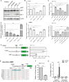
LMP1 induces activation of the mTORC1 signaling pathway. (A) HONE1-pLpcx and HONE1-LMP1 cells were lysed and analyzed by Western blotting using antibodies against LMP1 and various proteins in involved in mTORC1 signaling. (B) Various doses of 2117-LMP1 were transfected into HONE1 and 293T cells for 36 h and then examined by Western blotting to analyze for activation of the mTORC1 signaling pathway with specific antibodies. β-Actin expression was used as the loading control. (C) EBV-infected NPC cell line HONE1-M81 was infected with sh-LMP1 and control empty retroviral vector. The cellular RNA was extracted for qPCR analysis, and cell lysates were examined by Western blotting using antibodies against proteins involved in LMP1 and mTORC1 signaling pathways. Data are means ± SDs of triplicate measurements. **, P < 0.01.
FIG 2
FIG 2
LMP1 induces the expression of Glut-1 and increases glucose uptake. (A) NP69 and HONE1 cells were transfected with pcDNA and 2117-LMP1 expression plasmid. RNA was extracted 36 h later for RT-PCR analysis for Glut-1 to -4 gene transcription. GAPDH, glyceraldehyde-3-phosphate dehydrogenase. (B) Cells were lysed and the cell lysates were analyzed by Western blotting for LMP1 and Glut-1 expression using specific antibodies. β-Actin expression was used as the loading control. (C) RNA from NP460 and NP550 cells stably expressing LMP1 were extracted and analyzed by qPCR with Glut-1 primer. (D and E) The glucose consumption (D) and lactate production (E) of LMP1-transfected and control cells were determined. (F) Immunohistochemical staining revealed LMP1 and Glut-1 in formalin-fixed, paraffin-embedded NPC tissue sections. The images are of two representative NPCs. Images were acquired at a magnification of ×400. (G) Dot blot graph showing the immunoactivity scores of Glut-1 staining in NPC tumors with and without LMP1 expression. The median values of each group are shown by horizontal lines. Data are means ± SDs. *, P < 0.05; **, P < 0.01; ***, P < 0.005.
FIG 3
FIG 3
LMP1-induced NF-κB signaling activation is dependent on mTORC1. (A) HONE1-LMP1 cells were infected with sh-pScramble, sh-p65, and sh-IκBα lentivirus. Forty-eight hours after infection, cells were harvested and the cell lysates were analyzed by Western blotting for expression of LMP1 and mTORC1 pathway proteins with their corresponding antibodies. (B and C) HONE-LMP1 cells were treated with rapamycin for 24 h (B) and shRaptor for 48 h (C) and analyzed by Western blotting for expression of LMP1 and mTORC1 proteins. β-Actin expression was used as the loading control. (D) Cells were treated with 100 nM rapamycin for 24 h and analyzed by Western blotting using specific antibodies. (E) Cells were fixed and stained with p65-specific antibody and observed under a fluorescence microscope. (F) Relative luciferase reporter activity for NF-κB activation in LMP1-expressing cells was evaluated after various treatments. Data are means ± SDs of triplicate measurements. *, P < 0.05; **, P < 0.01; ***, P < 0.005.
FIG 4
FIG 4
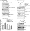
LMP1 upregulates Glut-1 transcription through the activation of mTORC1-mediated NF-κB signaling. (A) HONE1 cells stably expressing LMP1 were infected with different lentiviruses expressing shRNA against raptor, p65, and I-κB and their control scramble shRNA vector for 48 h. The cellular protein was subjected to Western blot analysis and the RNA to qPCR analysis. (B) Glucose consumption and lactate production in LMP1-expressing HONE1 cells after various treatments were determined. (C) Relative luciferase reporter activity of NF-κB was evaluated after various treatments. (D) Luciferase reporter vectors used to measure Glut-1 promoter activity. 293T cells were cotransfected with LMP1 and different reporter plasmids and the luciferase activity was determined 48 h later. (E) Motif analysis of p65 binding sites in selected Glut-1 promoter sequences. Multiple-sequence alignment among different species revealed a consensus sequence for NF-κB binding. (F) 293T cells were cotransfected with LMP1 plus pGL3-Glut1 (#1) or pGL3-Glut3-MUT (#4) reporter plasmid. The luciferase activity was measured after 48 h. (G) HONE1 cells were transfected with pcDNA and LMP1 expression plasmids. The cell lysates were subjected to ChIP assay using an anti-p65 antibody. Normal rabbit IgG antibody served as the negative control. PCR was performed to amplify a region surrounding the putative NF-κB binding region and a nonspecific NF-κB binding region. Data are means ± SDs of triplicate measurements. *, P < 0.05; **, P < 0.01; ***, P < 0.005.
FIG 5
FIG 5
Upregulation of Glut-1 mediates LMP1-induced cell proliferation and tumorigenesis. (A) NP69 cells were transfected with LMP1 expression plasmid and treated with STF-31 or infected with lentivirus expressing shGlut-1 for 48 h. The cells were then lysed and subjected to Western blotting using specific antibodies against Glut-1 and cleaved caspase 3. β-Actin expression was used as the loading control. (B) Cells were treated with STF-31 or infected with lentivirus expressing shGlut-1 for different time points and then assayed for [3H]thymidine uptake. (C and D) Glucose consumption (C) and lactate production (D) of NP69 cells after the indicated treatments were determined. (E and F) Representative results of colony formation (E) and anchorage-independent growth in soft agar (F) after various treatments. Histograms indicate the number of colonies formed and degree of anchorage-independent cell growth. Data are means ± SDs of triplicate measurements. *, P < 0.05; **, P < 0.01; ***, P < 0.005.
FIG 6
FIG 6
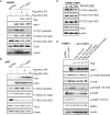
LMP1-induced mTORC1 signaling activation via the AKT/ERK/IKK signaling axis. (A) HONE1 and 293T cells were transiently transfected with LMP1 and examined after 36 h. The cells were lysed and subjected to Western blot analyses for activation of multiple signaling pathways as indicated. (B) HONE1 cells stably expressing LMP1 (HONE1-LMP1) were treated with U0126, LY294002, and rapamycin for 24 h. After that, an [3H]thymidine uptake assay was performed to estimate cell proliferation. (C) HONE1 cells were transfected with LMP1 expression vector and treated with U0126, NBD, and rapamycin for 48 h. After treatment, the cells were collected, lysed, and subjected to Western blot analyses for expression of LMP1 and mTORC1 pathway-related proteins. β-Actin expression was used as the loading control. (D) HONE1 cells transiently transfected with LMP1 cells either treated with rapamycin or infected with lentivirus expressing shRNA against Raptor were examined after 48 h. The cells were lysed and examined by Western blotting for IKK proteins using specific antibodies. β-Actin expression was used as the loading control.
FIG 7
FIG 7
C-terminal activating regions (CTARs) of LMP1 are involved in LMP1-induced mTORC1 activation. (A) Schematic illustrations of wild-type LMP1 and its mutants used for thymidine incorporation, mTORC1 activation, and glucose metabolism. WT, wild-type LMP1; 3A, LMP1 harboring CTAR1 mutation; 8C, LMP1 harboring CTAR2 deletion; 3A/8C, LMP1 harboring CTAR1 mutation and deletion of CTAR2. (B) HONE1 cells were transfected with different LMP1 constructs and examined for [3H]thymidine uptake, which reflects cell proliferation. (C) HONE1 and 293T cells were transfected with different types of LMP1 constructs. The cells were lysed and analyzed by Western blotting for LMP1 and mTORC1 proteins. (D) HONE1 cells were transfected with different types of LMP1 constructs and then analyzed for activation of the AKT, MEK/ERK, and IKK pathways using specific antibodies. β-Actin was used as the loading control. (E to H) HONE1 cells were transfected with different LMP1 constructs and analyzed for protein expression of Glut-1(E), RNA levels of Glut-1 (F), glucose consumption (G), and lactate production (H) after transfection with wild-type and mutant LMP1s. Data are means ± SDs of triplicate measurements. *, P < 0.05; **, P < 0.01; ***, P < 0.005.
FIG 8
FIG 8
IKKβ is the main contributor to LMP1-induced mTORC1 activation. (A) HONE1 cells were cotransfected with LMP1 and Flag-IKKα and -β KM mutants for 36 h and then analyzed by Western blotting for activation of the NF-κB and mTORC1 pathways. β-Actin expression was used as the loading control. (B) HONE1-LMP1 cells were treated with PS1145 for 24 h and then analyzed by Western blotting for detection of the mTORC1 and NF-κB pathways. (C and D) HONE1 cells were transfected with different IKK mutant constructs and then examined for expression of Glut-1 protein by Western blotting (C) and Glut-1 mRNA transcription by qPCR (D). (E to H) Glucose consumption (E and G) and lactate production (F and H) were measured after the indicated treatments. Data are means ± SDs of triplicate measurements. *, P < 0.05; **, P < 0.01.
FIG 9
FIG 9
IKKβ phosphorylates TSC2 at Ser939 to activate mTORC1. (A and B) HONE1 (A) and 293T (B) cells were cotransfected with LMP1 and wild-type IKKβ (Flag-IKKβ WT) and mutant IKKβ (Flag-IKKβ-KM) constructs for 36 h. Different phosphorylation sites of TSC2 and other relevant proteins were then analyzed with corresponding antibodies in transfected cells. β-Actin expression was used as the loading control. (C) HONE1-LMP1 cells were treated with PS1145 for 24 h and then analyzed by Western blotting for detection of the different phosphorylation sites of TSC2 by the specific antibodies. (D) HONE1 cells were cotransfected with different phosphorylation site-mutated TSC2 plasmids and 2117-LMP1 for 36 h. The cells were collected and cell lysates were subjected to Western blot analyses. The phosphorylation of mTORC1 components was used to monitor mTORC1 signaling activity. β-Actin expression was used as the loading control.
FIG 10
FIG 10
Schematic diagram of LMP1-induced mTORC1/NF-κB/Glut-1 activation in NPC cells. The viral oncoprotein LMP1 activates mTORC1 through AKT/ERK/IKK signaling axis. Both CTAR1 and CTAR2 of LMP1 are involved in activation of mTORC1 signaling. LMP1-CTAR1 regulates mTORC1 activation through AKT-ERK-IKKα, while LMP1-CTAR2 regulates mTROC1 activation via IKKβ. The CTAR2/IKKβ plays a more dominant role in mTORC1 activation through phosphorylation of Ser939 of TSC2. Furthermore, mTORC1 activation induced by LMP1 also modulates NF-κB signaling by undefined pathways (dotted arrows), which regulates aerobic glycolysis and promotes tumorigenesis through upregulation of Glut-1 transcription and expression.

Similar articles

Cited by

References

    1. Strong JE, Coffey MC, Tang D, Sabinin P, Lee PW. 1998. The molecular basis of viral oncolysis: usurpation of the Ras signaling pathway by reovirus. EMBO J 17:3351–3362. doi:10.1093/emboj/17.12.3351. - DOI - PMC - PubMed
    1. Wei WI, Sham JS. 2005. Nasopharyngeal carcinoma. Lancet 365:2041–2054. doi:10.1016/S0140-6736(05)66698-6. - DOI - PubMed
    1. Cheng SH, Tsai SY, Yen KL, Jian JJ-M, Chu N-M, Chan K-Y, Tan T-D, Cheng JC, Hsieh C-Y, Huang AT. 2000. Concomitant radiotherapy and chemotherapy for early-stage nasopharyngeal carcinoma. J Clin Oncol 18:2040–2045. - PubMed
    1. Pathmanathan R, Prasad U, Sadler R, Flynn K, Raabtraub N. 1995. Clonal proliferations of cells infected with Epstein-Barr virus in preinvasive lesions related to nasopharyngeal carcinoma. N Engl J Med 333:693–698. doi:10.1056/NEJM199509143331103. - DOI - PubMed
    1. Trivedi P, Hu LF, Chen F, Masucci M, Klein G, Winberg G, Christensson B. 1994. Epstein-Barr virus (EBV)-encoded membrane protein LMP1 from a nasopharyngeal carcinoma is non-immunogenic in a murine model system, in contrast to a B cell-derived homologue. Eur J Cancer 30:84–88. doi:10.1016/S0959-8049(05)80024-3. - DOI - PubMed

MeSH terms

Substances

LinkOut - more resources