. 2017 Jan 5:7:40042.
doi: 10.1038/srep40042.

Evaluation of left pulmonary artery sling, associated cardiovascular anomalies, and surgical outcomes using cardiovascular computed tomography angiography

Affiliations

Evaluation of left pulmonary artery sling, associated cardiovascular anomalies, and surgical outcomes using cardiovascular computed tomography angiography

Jiajun Xie et al. Sci Rep. .

Abstract

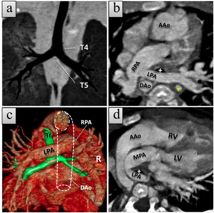
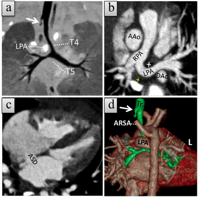
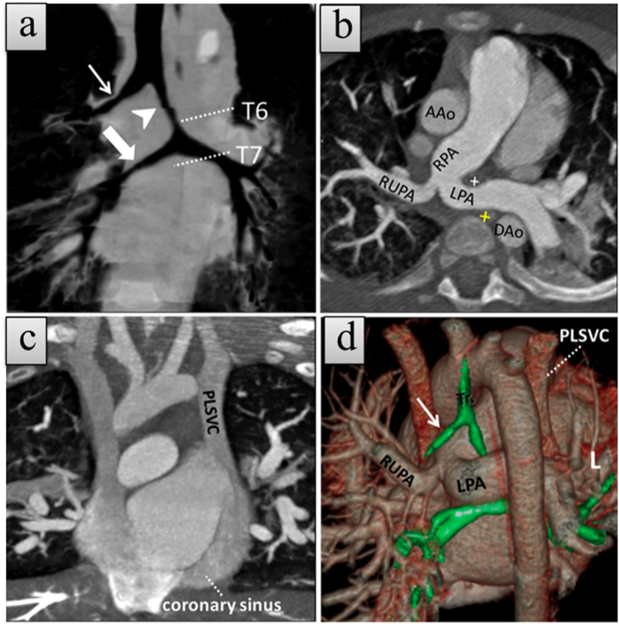
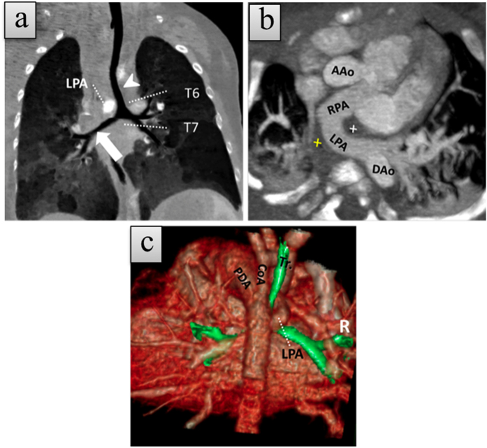
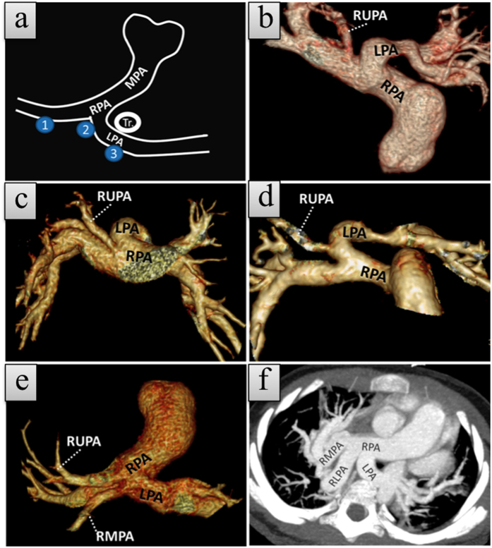
We evaluated the prevalence, image appearance, associated cardiovascular anomalies, and surgical outcomes of left pulmonary artery sling (LPAS) using cardiovascular computed tomography angiography (CCTA). A retrospective search of patients from our database between October 2007 and December 2014 identified 52,200 patients with congenital heart diseases (CHD) referred for CCTA, echocardiography, or magnetic resonance imaging. Clinical information, CCTA findings, associated cardiovascular anomalies, and surgical outcomes were analyzed. We showed a hospital-based prevalence of 71 patients with LPAS (0.14%, 71/52,200) among CHD patients. Of these, 47 patients with CCTA examinations were assessed further. Most patients (40/47, 85%) had associated cardiovascular anomalies, of which ventricular septal defects (22/47, 47%), atrial septal defects (20/47, 43%), patent ductus arteriosus (16/47, 34%), persistent left superior vena cava (14/47, 30%), and abnormal branching of the right pulmonary artery (ABRPA) (14/47, 30%) were most commonly identified. In total, 28 patients underwent LPA reanastomosis and/or tracheoplasty in our center, and 5 died. LPAS had a hospital-based prevalence of 0.14% among CHD patients. ABRPA is not uncommon and must be recognized. CCTA is a feasible method for demonstrating LPAS and its associated cardiovascular anomalies for an optimal pre-operative assessment of LPAS.


Figures

Figure 1
Figure 1. A 1-month-old girl with type 1A left pulmonary artery sling (LPAS) with dextro-transposition of the great arteries (d-TGA).
(a) Coronal minimum-intensity projection (MinIP) CCTA image shows a normal tracheobronchial branch pattern. (b, c) Axial thin slice maximum-intensity projection (MIP) and posterior view of merged volume render CCTA images show anomalous left pulmonary artery (LPA) rising from the proximal right pulmonary artery (RPA) and coursing between the trachea (white cross in b and Tr in c) and esophagus (cross in yellow). The esophagus lumen showed a high density due to presence of a nasogastric tube. (d) Oblique thin slice MIP CCTA image demonstrates ascending aorta (AAo) rising from the right ventricle (RV) and the main pulmonary artery (MPA) from the left ventricle (LV), indicating the d-TGA. Luminal dot line = descending aorta. The patient also has an atrial septal defect and ventricular septal defect (not shown).
Figure 2
Figure 2. A 9-month-old girl with type 1B left pulmonary artery sling (LPAS) with atrial septal defect (ASD) and aberrant right subclavian artery (ARSA).
(a) Coronal minimum-intensity projection (MinIP) CCTA image shows a normal level of bronchial bifurcation but with a right rudimentary tracheal bronchus (white arrow) and an abnormal course of the left pulmonary artery (LPA). (b,c), Axial thin slice maximum-intensity projection (MIP) CCTA images demonstrate ASD and anomalous LPA originating from the proximal right pulmonary artery (RPA) and coursing between the trachea (cross in white) and esophagus (cross in yellow). (d) Posterior view of the trachea (Tr), and cardiovascular merged volume render CCTA image reveals rudimentary right rudimentary tracheal bronchus (white arrow) and ARSA arising from the aortic isthmus. The patient also has a ventricle septal defect (not shown).
Figure 3
Figure 3. A 6-month-old boy with type 2A left pulmonary artery sling (LPAS) with persistent left superior vena cava (PLSVC).
(a) Coronal minimum-intensity projection (MinIP) CCTA image shows a lower level of bronchial bifurcation with a classic tracheal bronchus (white arrow), left intermediate bronchus (arrowhead), and bridging bronchus (broad arrow). (b) Axial thin slice maximum-intensity projection (MIP) CCTA image shows anomalous left pulmonary artery (LPA) arising from the proximal right pulmonary artery (RPA) and coursing between the trachea (cross in white) and esophagus (cross in yellow). The esophagus lumen showed a high density due to a nasogastric tube. Right upper pulmonary artery (RUPA) can be seen originating from the common orifice of LPA and RPA; (c) Coronal thin slice MIP CCTA image demonstrates PLSVC draining into the right atrium via the coronary sinus. (d) Posterior view of trachea (Tr) and cardiovascular merged volume render CCTA image shows tracheal bronchus (white arrow) and anomalous LPA course. The patient also has atrial septal defect and ventricular septal defects (not shown).
Figure 4
Figure 4. 1-month-old girl with type 2B left pulmonary artery sling (LPAS) with coarctation of aorta (CoA) and patent ductus arteriosus (PDA).
(a) Coronal thin slice minimum-intensity projection (MinIP) CCTA image shows a lower level of bronchial bifurcation with classic left intermediate bronchus (arrowhead) and bridging bronchus (broad arrow). (b) Axial thin slice maximum-intensity projection (MIP) CCTA image reveals anomalous LPA originating from the proximal RPA and coursing between the trachea (cross in white) and esophagus (cross in yellow). (c) Volume render CCTA image shows an abnormal course of the LPA, a big PDA, and CoA.
Figure 5
Figure 5. Volume rendering (B–E) and maximum-intensity-projection (MIP, F) images show normal and anomalous origin of the proximal branches of the right pulmonary artery (RPA) in five patients with left pulmonary artery sling.
(a) Schematic diagram shows the origin sites of proximal right pulmonary artery branches: RPA, common orifice, left pulmonary artery (LPA). (b–e) Volume-rendered images show the origins of the proximal branches of the RPA in our series. (b) RUPA originates from the RPA, (c) RUPA originates from the common orifice of the LPA and RPA, (d) RUPA originates from the LPA, (e) RMPA originates from the LPA and inferior to the RUPA origin site, and (f) MIP image shows RLPA originating from the common orifice of the LPA and RPA.

Similar articles

Cited by

References

    1. Pawade A., de Leval M. R., Elliott M. J. & Stark J. Pulmonary artery sling. Ann Thorac Surg 54, 967–970 (1992). - PubMed
    1. Wells T. R., Gwinn J. L., Landing B. H. & Stanley P. Reconsideration of the anatomy of sling left pulmonary artery: the association of one form with bridging bronchus and imperforate anus. Anatomic and diagnostic aspects. J Pediatr Surg 23, 892–898 (1988). - PubMed
    1. Gikonyo B. M., Jue K. L. & Edwards J. E. Pulmonary vascular sling: report of seven cases and review of the literature. Pediatr Cardiol 10, 81–89, doi: 10.1007/BF02309919 (1989). - DOI - PubMed
    1. Leonardi B. et al.. Imaging modalities in children with vascular ring and pulmonary artery sling. Pediatr Pulmonol 50, 781–788, doi: 10.1002/ppul.23075 (2015). - DOI - PubMed
    1. Hu X. H., Pa M. E., Shen Q. L. & Huang G. Y. Multi-detector computed tomography evaluation of tracheobronchial anomaly in pediatric patients with left pulmonary artery sling. Chin Med J(Engl) 126, 2790–2792 (2013). - PubMed

Publication types