Review
. 2016 Dec 20:12:73-86.
doi: 10.2147/IJN.S117501. eCollection 2017.

Self-assembled peptide nanomaterials for biomedical applications: promises and pitfalls

Affiliations
Review

Self-assembled peptide nanomaterials for biomedical applications: promises and pitfalls

Linlin Sun et al. Int J Nanomedicine. .

Abstract

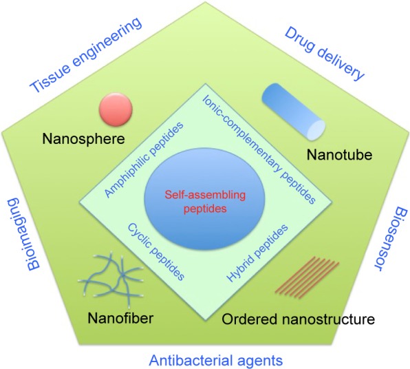
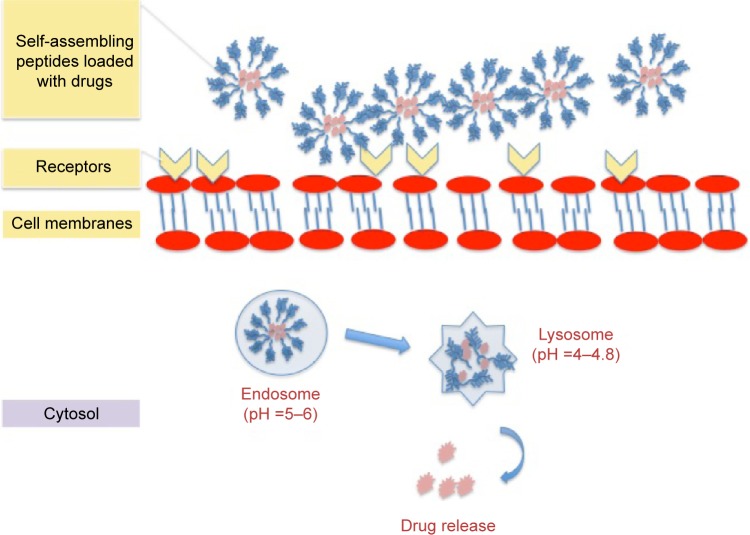
Over the last several decades, a great number of advances have been made in the area of self-assembled supramolecules for regenerative medicine. Such advances have involved the design, preparation, and characterization of brand new self-assembled peptide nanomaterials for a variety of applications. Among all biomolecules considered for self-assembly applications, peptides have attracted a great deal of attention as building blocks for bottom-up fabrication, due to their versatility, ease of manufacturing, low costs, tunable structures, and versatile properties. Herein, some of the more exciting new designs of self-assembled peptides and their associated unique features are reviewed and several promising applications of how self-assembled peptides are advancing drug delivery, tissue engineering, antibacterial therapy, and biosensor device applications are highlighted.

Keywords: antibacterial therapy; biomedical applications; biosensor devices; drug delivery; peptides; self-assembly.


Conflict of interest statement

The authors report no conflicts of interest in this work.

Figures

Figure 1
Figure 1
Schematic illustration of various nanostructures (nanosphere, nanotube, nanofiber, and ordered nanostructure) formed by self-assembling peptides (amphiphilic peptides, ionic-complementary peptides, cyclic peptides, and hybrid peptides) and their applications in tissue engineering, drug delivery, bioimaging, and biosensors.
Figure 2
Figure 2
Different types of ionic-complementary peptides. Notes: (A) The hydrophobic side of the peptide molecule is green, negative charges are red, and positive charges are blue. (a) The peptide structure has one hydrophobic side and one side with negative charges. (b) The peptide structure has one hydrophobic side and one side with positive charges. (c) The peptide structure has one hydrophobic side and one side with alternate positive and negative charges. (B) Four representative ionic peptides: RAD16-I, RAD16-II, EAK16-I, and EAK16-II. Reprinted from Nano Today, Volume 4, Edition 2, Yang Y, Khoe U, Wang X, Horii A, Yokoi H, Zhang S. Designer self-assembling peptide nanomaterials, pages 193–210, Copyright 2009 with permission from Elsevier.
Figure 3
Figure 3
(A) Molecular structure of an alkylated peptide with four regions: hydrophobic tail, beta-sheet forming segment, charged groups, and bioactive epitope. Adapted from Cui H, Webber MJ, Stupp SI. Self-assembly of peptide amphiphiles: from molecules to nanostructures to biomaterials. Biopolymers. 2010;94(1):1–18. Copyright © 2010 Wiley Periodicals, Inc. (B) Schematic illustration of an alkylated peptide (IKVAV) molecule and the nanofiber formed by IKVAV peptides. (C) TEM image of IKVAV nanofibers. From Silva GA, Czeisler C, Niece KL, et al. Selective differentiation of neural progenitor cells by high-epitope density nanofibers. Science. 2004;303(5662):1352–1355. Reprinted with permission from AAAS. Abbreviation: TEM, transmission electron microscopy.
Figure 4
Figure 4
Schematic illustration of the cyclic peptide molecule and the nanotubular structure provided by cyclic peptides. Notes: Reprinted with permission from Macmillan Publishers Ltd: Nature. Fernandez-Lopez S, Kim HS, Choi EC, et al. Antibacterial agents based on the cyclic D,L-alpha-peptide architecture. 2001;412(6845):452–455.
Figure 5
Figure 5
Schematic illustration of cell-membrane interactions and drug release of the drug-loaded self-assembling peptides.
Figure 6
Figure 6
(A) Schematic illustration of paclitaxel-loaded Tat peptide nanofibers. (B) TEM image of paclitaxel-loaded Tat nanofibers in PBS. (C) Confocal image of the endocytosis pathway of drug release. (D) CD spectrum of the Tat peptide nanofibers. (E) KB-3-1 cervical cancer cell inhibition by the drug-loaded peptides. Reprinted with permission from Zhang P, Cheetham AG, Lin YA, Cui H. Self-assembled tat nanofibers as effective drug carrier and transporter. Acs Nano. 2013;7(7):5965–5977. Copyright 2013 American Chemical Society. Abbreviations: TEM, transmission electron microscopy; PBS, phosphate-buffered saline; CD, circular dichroism.
Figure 7
Figure 7
(A) After heating, amphiphilic peptides formed the hydrogel in a PBS solution. (B) After mixing with a CaCl2 solution, the peptide solution formed a string-like hydrogel. (C) Mesenchymal stem cells grew and differentiated at along the direction aligned with the hydrogel string formed from the peptides. (D) Fluorescence image of calcein-labeled human mesenchymal stem cells in the peptide hydrogel. Reprinted by permission from Macmillan Publishers Ltd: Nature Materials. Zhang S, Greenfield MA, Mata A, et al. A self-assembly pathway to aligned monodomain gels. 2010;9(7):594–601., Copyright 2010. Abbreviation: PBS, phosphate-buffered saline.
Figure 8
Figure 8
CG-MD images of the interaction of self-assembling cyclic peptides ([RRKWLWLW]) with POPE–POPG lipid membranes. Notes: (A) With a ratio of 1/22 CP/Lip, the upper image is the initial interaction at 0 ns and the lower image is the final simulation omitting the peptides at 165 ns. (B) With the ratio of 1/10 CP/Lip at 658 ns, the top image is the top view, the middle image is the side view of the initial interaction, and the lower image is the final simulation omitting the peptides. (C) With the ratio of 1/7 CP/Lip at 1.02 μs, the top image is the top view, the middle image is the side view of the initial interaction, and the lower image is the final simulation omitting the peptides. (D) With the ratio of 1/5 CP/Lip at 6.12 μs, the top image is the top view, the middle image is the side view of the initial interaction, and the lower image is the final simulation omitting the peptides. Reprinted with permission from Khalfa A, Tarek M. On the antibacterial action of cyclic peptides: insights from coarse-grained MD simulations. J Phys Chem. 2010;114(8):2676–2684. Copyright 2010 American Chemical Society. Abbreviations: CG-MD, coarse-grained molecular dynamic; POPE, palmitoyl-oleoyl-phosphatidylethanolamine; POPG, palmitoyl-oleoyl-phosphatidylglycerol; CP/Lip, cyclic peptide/lipid.
Figure 9
Figure 9
(A) The molecular structure of the pyrene-labeled peptide amphiphiles 1 and 2. (B) Schematic representation of the fluorescence light off and light up detections to copper and silver ions. Reproduced from Kim I, Jeong HH, Kim YJ, et al. A “light-up” 1D supramolecular nanoprobe for silver ions based on assembly of pyrene-labeled peptide amphiphiles: cell-imaging and antimicrobial activity. J Mat Chem. 2014;2(38):6478–6486, with permission of The Royal Society of Chemistry. Abbreviation: PET, photoinduced electron transfer.

References

    1. Whitesides GM, Boncheva M. Beyond molecules: self-assembly of mesoscopic and macroscopic components. Proc Natl Acad Sci U S A. 2002;99(8):4769–4774. - PMC - PubMed
    1. Whitesides GM, Grzybowski B. Self-assembly at all scales. Science. 2002;295(5564):2418–2421. - PubMed
    1. Tu RS, Tirrell M. Bottom-up design of biomimetic assemblies. Adv Drug Deliv Rev. 2004;56(11):1537–1563. - PubMed
    1. Feynman RP. There’s plenty of room at the bottom. Caltech Eng Sci. 1959;23(5):22–36.
    1. Lehn JM. Supramolecular chemistry – scope and perspectives molecules, supermolecules, and molecular devices. Angew Chem Int Ed Engl. 1988;27(1):89–112.

LinkOut - more resources