. 2016 Dec 20:12:127-135.
doi: 10.2147/IJN.S107491. eCollection 2017.

Bottom-up fabrication of zwitterionic polymer brushes on intraocular lens for improved biocompatibility

Affiliations

Bottom-up fabrication of zwitterionic polymer brushes on intraocular lens for improved biocompatibility

Yuemei Han et al. Int J Nanomedicine. .

Abstract

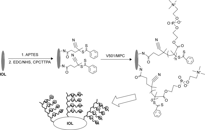
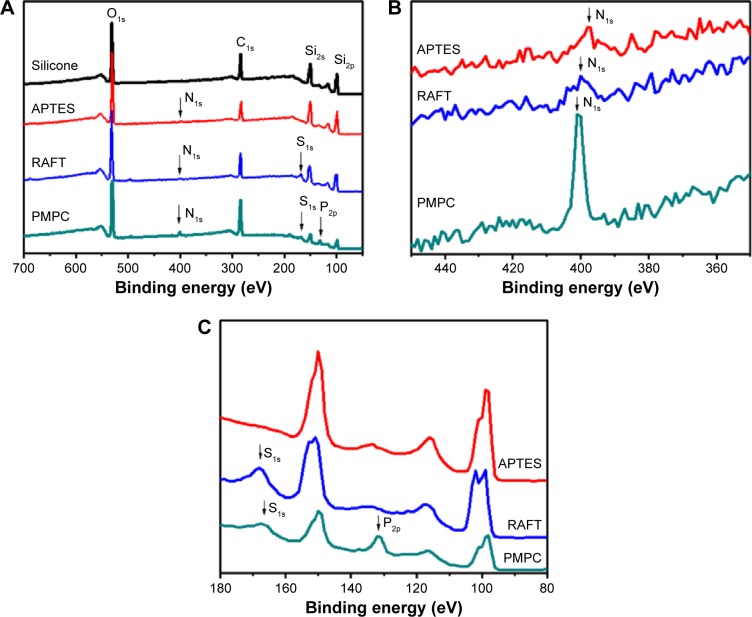
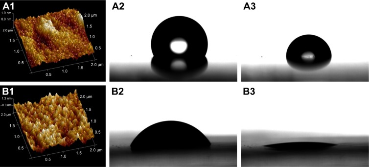
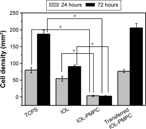
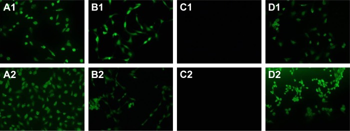
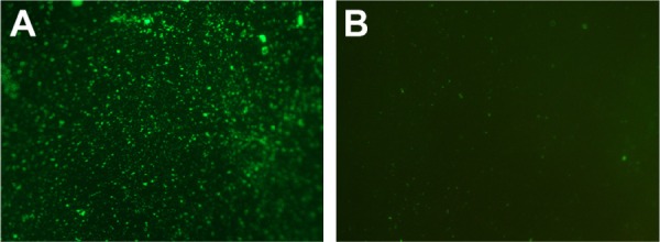
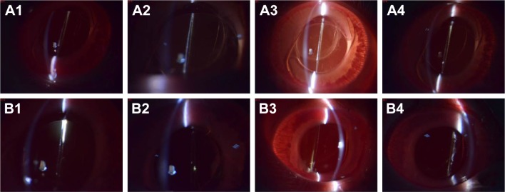
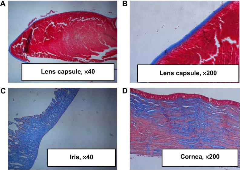
Intraocular lens (IOL) is an efficient implantable device commonly used for treating cataracts. However, bioadhesion of bacteria or residual lens epithelial cells on the IOL surface after surgery causes postoperative complications, such as endophthalmitis or posterior capsular opacification, and leads to loss of sight again. In the present study, zwitterionic polymer brushes were fabricated on the IOL surface via bottom-up grafting procedure. The attenuated total reflection-Fourier transform infrared and contact angle measurements indicated successful surface modification, as well as excellent hydrophilicity. The coating of hydrophilic zwitterionic polymer effectively decreased the bioadhesion of lens epithelial cells or bacteria. In vivo intraocular implantation results showed good in vivo biocompatibility of zwitterionic IOL and its effectiveness against postoperative complications.

Keywords: PCO; RAFT; endophthalmitis; in vivo; surface modification.


Conflict of interest statement

The authors report no conflicts of interest in this work.

Figures

Figure 1
Figure 1
Schematic illustration of surface-initiated RAFT polymerization of MPC on IOL surface. Abbreviations: RAFT, reversible addition-fragmentation chain-transfer; MPC, 2-methacryloyloxyethyl phosphorylcholine; IOL, intraocular lens; APTES, (3-aminopropyl) triethoxysilane; EDC, N-(3-dimethylaminopropyl)-N′-ethylcarbodiimide hydrochloride; NHS, N-hydroxysulfosuccinimide sodium; CPCTTPA, 4-cyano-4-(phenyl-carbonothioylthio) pentanoic acid; V501, 2-morpholino-ethanesulfonic acid, 4,4′-azobis-(4-cyanovaleric acid).
Figure 2
Figure 2
XPS pattern of silicone IOL materials and materials after APTES aminolysis or RAFT agent immobilization, followed by SI-RAFT polymerization of MPC. Notes: (A) The full XPS pattern. (B) The amplified view of the pattern in 350–450 eV. (C) The amplified view of the pattern in 80–180 eV. Abbreviations: XPS, X-ray photoelectron spectroscopy; IOL, intraocular lens; APTES, (3-aminopropyl) triethoxysilane; RAFT, reversible addition-fragmentation chain-transfer; SI-RAFT, surface initiated RAFT; MPC, 2-methacryloyloxyethyl phosphorylcholine; PMPC, poly(MPC).
Figure 3
Figure 3
The AFM images (A1 and B1) and water contact angle of silicone IOL materials before (A1A3) and after (B1B3) PMPC modification. The contact angle photos were taken immediately after the water drop contacted materials (A2 and B2) and after 480 seconds (A3 and B3). Abbreviations: AFM, atomic force microscopy; IOL, intraocular lens; PMPC, poly(2-methacryloyloxyethyl phosphorylcholine).
Figure 4
Figure 4
Cell densities on different surfaces after culturing for 24 and 72 hours. Notes: Transferred IOL-PMPC refers to that the cells were originally cultured on IOL-PMPC plate for 24 hours and then the culture supernatant was transferred onto TCPS. *Indicates P<0.05. Abbreviations: IOL, intraocular lens; PMPC, poly(2-methacryloyloxyethyl phosphorylcholine); TCPS, tissue culture polystyrene.
Figure 5
Figure 5
The representative fluorescent images of the LECs cultured on surface of different materials for (A1D1) 24 and (A2D2) 72 hours. (A1 and A2) TCPS. (B1 and B2) Silicone IOL materials. (C1 and C2) IOL materials surface coated with PMPC brushes. (D1 and D2) Cells were originally cultured on IOL-PMPC plate for 24 hours, and then the culture supernatant was transferred onto TCPS. The magnification is ×200. Abbreviations: LECs, lens epithelial cells; TCPS, tissue culture polystyrene; IOL, intraocular lens; PMPC, poly(2-methacryloyloxyethyl phosphorylcholine).
Figure 6
Figure 6
Fluorescence microscopy images of Staphylococcus aureus adhesions on (A) pristine silicone IOL material and (B) PMPC brush-modified silicone IOL material. The magnification is ×100. Abbreviations: IOL, intraocular lens; PMPC, poly(2-methacryloyloxyethyl phosphorylcholine).
Figure 7
Figure 7
Representative slit lamp images of the (A1A4) implanted PMPC-modified hydrophobic IOL and (B1B4) the implanted pristine hydrophobic IOL taken on days 1, 3, 7, and 30. The magnification is ×16. Abbreviations: PMPC, poly(2-methacryloyloxyethyl phosphorylcholine); IOL, intraocular lens.
Figure 8
Figure 8
The histochemical stain of the PMPC brushes modified hydrophobic IOL implantation. (A, B) The lens capsule; (C) iris; (D) cornea. Abbreviations: PMPC, poly(2-methacryloyloxyethyl phosphorylcholine); IOL, intraocular lens.

Similar articles

Cited by

References

    1. Nibourg LM, Gelens E, Kuijer R, Hooymans JMM, van Kooten TG, Koopmans SA. Prevention of posterior capsular opacification. Exp Eye Res. 2015;136:100–115. - PubMed
    1. Saika S, Kawashima Y, Miyamoto T, et al. Pathological findings in lens capsule and silicone intraocular lens extracted from eye with chronic infectious endophthalmitis. JNP J Ophthalmol. 1999;42(6):456–460. - PubMed
    1. Okajima Y. Biofilm formation by Staphylococcus epidermidis on intraocular lens material. Invest Ophthalmol Vis Sci. 2006;47(7):2971–2975. - PubMed
    1. Lin QK, Ren KF, Ji J. Hyaluronic acid and chitosan-DNA complex multilayered thin film as surface-mediated nonviral gene delivery system. Colloids Surf B Biointerfaces. 2009;74(1):298–303. - PubMed
    1. Lin QK, Ding X, Qiu FY, Song XX, Fu GS, Ji J. In situ endothelialization of intravascular stents coated with an anti-CD34 antibody functionalized heparin-collagen multilayer. Biomaterials. 2010;31(14):4017–4025. - PubMed