. 2016:2016:3718468.
doi: 10.1155/2016/3718468. Epub 2016 Dec 8.

Macroautophagy and Selective Mitophagy Ameliorate Chondrogenic Differentiation Potential in Adipose Stem Cells of Equine Metabolic Syndrome: New Findings in the Field of Progenitor Cells Differentiation

Affiliations

Macroautophagy and Selective Mitophagy Ameliorate Chondrogenic Differentiation Potential in Adipose Stem Cells of Equine Metabolic Syndrome: New Findings in the Field of Progenitor Cells Differentiation

Krzysztof Marycz et al. Oxid Med Cell Longev. 2016.

Erratum in

Abstract

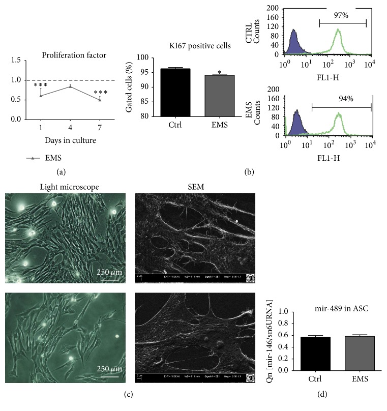
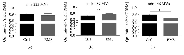
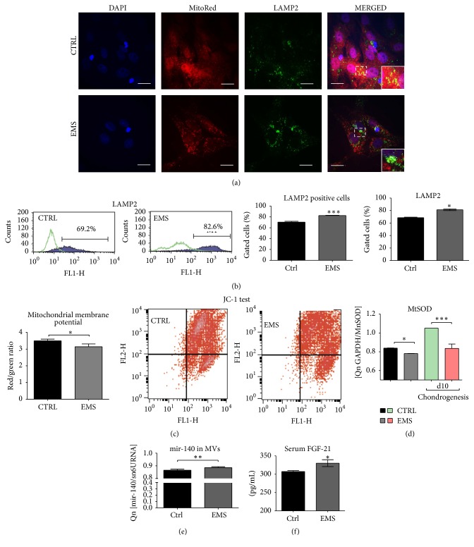
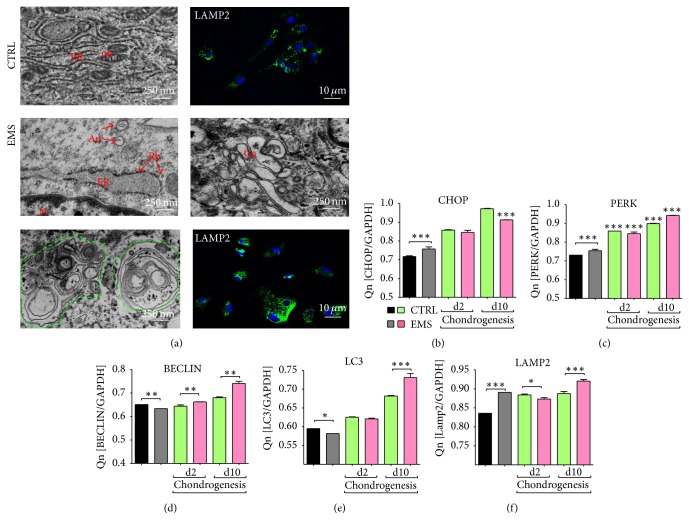
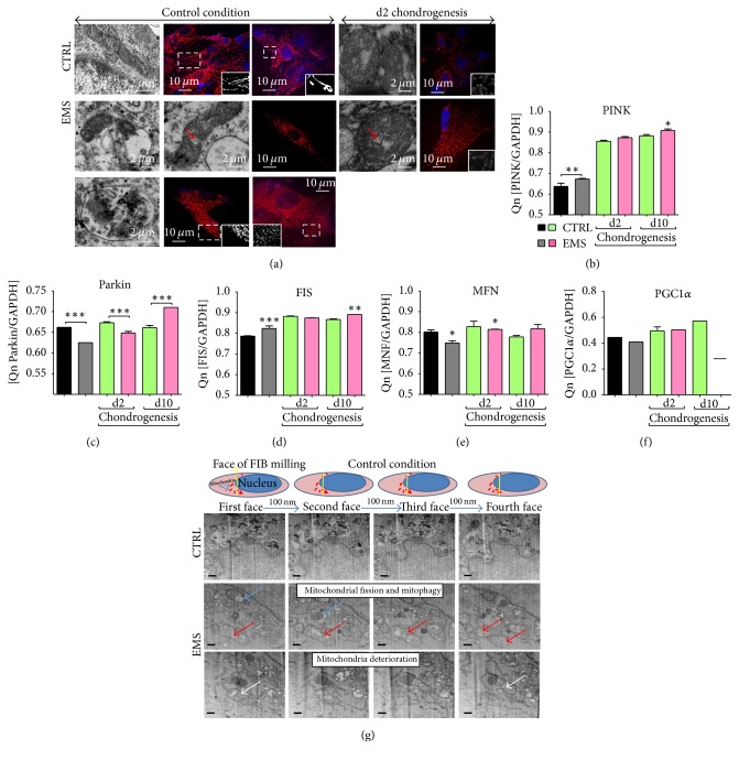
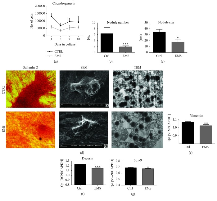
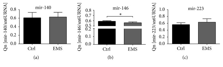
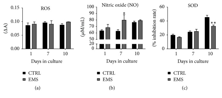
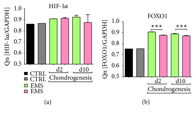
Equine metabolic syndrome (EMS) is mainly characterized by insulin resistance, obesity, and local or systemic inflammation. That unfriendly environment of adipose tissue has huge impact on stem cells population (ASC) residing within. In the present study, using molecular biology techniques and multiple imaging techniques (SEM, FIB-SEM, and confocal microscopy), we evaluated the impact of EMS on ASC viability and chondrogenic differentiation. Moreover, we visualized the mitochondrial network and dynamics in ASCCTRL and ASCEMS during control and chondrogenic conditions. In control conditions, ASCEMS were characterized by increased mitochondrial fission in comparison to ASCCTRL. We found that extensive remodeling of mitochondrial network including fusion and fission occurs during early step of differentiation. Moreover, we observed mitochondria morphology deterioration in ASCEMS. These conditions seem to cause autophagic shift in ASCEMS, as we observed increased accumulation of LAMP2 and formation of multiple autophagosomes in those cells, some of which contained dysfunctional mitochondria. "Autophagic" switch may be a rescue mechanism allowing ASCEMS to clear impaired by ROS proteins and mitochondria. Moreover it provides a precursors-to-macromolecules synthesis, especially during chondrogenesis. Our data indicates that autophagy in ASCEMS would be crucial for the quality control mechanisms and maintenance of cellular homeostasis ASCEMS allowing them to be in "stemness" status.


Conflict of interest statement

The authors declare that there is no conflict of interests.

Figures

Figure 1
Figure 1
Characterization of isolated ASC analysis of surface antigens and multipotency assay. Using flow cytometer, the expression of CD44, CD45, CD90, and CD105 antigens was investigated. Isolated cells were characterized by the expression of CD44, CD90, and CD105 while they lacked the expression of CD45 hematopoietic marker (a). Interestingly, ASCEMS displayed higher expression of CD44. Multipotency of ASC was confirmed by trilineage differentiation assay. Representative photographs showing the effectiveness of chondrogenesis (Safranin O), osteogenesis (Alizarin Red), and adipogenesis (LipidTox) (b). Cells cultured in standard culture medium served as a control.
Figure 2
Figure 2
Proliferation and morphology of ASC. Using resazurin-based assay, the growth kinetics of isolated cells was established. During the seven-day test ASCEMS displayed decreased proliferation potential in comparison to control group (a). Flow cytometry analysis confirmed decreased expression of Ki67 in those cells (b). Morphology of investigated cells was evaluated after day 7. While ASCCTRL reached high confluence and developed robust net of cytoskeletal projections (lamellipodia and filopodia), ASCEMS did not form multilayer and were more amorphous in shape with reduced cellular projections (c). Interestingly, there were no differences in the expression of mir-489, which plays crucial role in the differentiation process (d). Results are expressed as mean ± SD. ∗∗∗ p value < 0.001.
Figure 3
Figure 3
Evaluation of miRNA content in microvesicles (MVs) secreted by ASCCTRL and ASCEMS. There were no significant differences in the expression of mir-223 in investigated groups (a). Interestingly, secreted in MVs, mir-489 was upregulated in ASCEMS (b). On the contrary, mir-146, whose decreased expression is correlated with inflammation and diabetic wound healing, was downregulated in ASCEMS (c). Results are expressed as mean ± SD. p < 0.05, ∗∗ p < 0.01.
Figure 4
Figure 4
Mitochondria condition and clearance in ASCCTRL and ASCEMS. Representative photographs showing the results of DAPI, MitoRed, and anti-LAMP2 stainings. Interestingly, although LAMP2 expression was increased in ASCEMS, clearance of mitochondria in those cells seems to be reduced as we observed decreased number of mitochondria fused with lysosomes in comparison to control group (a). Flow cytometry analysis confirmed increased expression of LAMP2 in ASCEMS (b). Mitochondria deterioration was confirmed with flow cytometry using JC1 test, as we observed decreased mitochondrial membrane potential in ASCEMS (c). Moreover, the antioxidative protection coming from mitochondrial MnSOD was reduced (d) as established with qRT-PCR. The mir-140 transported via MVs was increased in ASCEMS (e) which correlates with increased expression of MFN. The serum level of FGF21, characteristic of obesity and diabetes, was upregulated in ASCEMS (f). Results are expressed as mean ± SD. p < 0.05, ∗∗ p < 0.01, and ∗∗∗ p < 0.001. Scale bars: 10 μm.
Figure 5
Figure 5
Endoplasmic reticulum (ER) stress and autophagy. The autophagy in investigated ASC was established using transmission and confocal microscopy (a). Both ER and Golgi compartments were grossly expanded in ASCEMS. Moreover, large part of ER become fragmented and disintegrated. Large double-membrane vacuoles, multilamellar autophagic bodies (green outline), preautophagosome structures, and engulfed organelles were characterized for ASCEMS. Moreover, anti-LAMP2 immunofluorescence staining revealed increased lysosome accumulation in those cells. The expression of ER stress-related genes—CHOP (b) and PERK (c)—was increased in ASCEMS in control conditions but during chondrogenic differentiation the amount of its mRNA was decreased in comparison to control group. Transcription of genes involved in the autophagy process including Beclin (d) and LC3 (e) in standard culture was decreased in ASCEMS, but during chondrogenesis it significantly increased. mRNA level for LAMP2 was upregulated in those cells in both standard and chondrogenic cultures (f) despite day 2. Results are expressed as mean ± SD. p < 0.05, ∗∗ p < 0.01, and ∗∗∗ p < 0.001. Au: autophagosomes, Er: endoplasmic reticulum, Rb: ribosomes, N: nucleus, Ga: Golgi apparatus, Mt: mitochondria.
Figure 6
Figure 6
Mitochondria dynamics in ASCCTRL and ASCEMS. Distribution of ASC mitochondria was evaluated using TEM and confocal microscopy with MitoRed staining (a). In control conditions ASCCTRL mitochondria presented typical, elongated, bean shape morphology with well-developed cristae. A long net of connected mitochondria was observed although, during chondrogenesis, fragmented mitochondrial phenotype was observed. In ASCEMS, mitochondrial fission was enhanced during both control and chondrogenic conditions. Using RT-PCR we evaluated the expression of PINK (b), PARKIN (c), FIS (d), MFN (e), and PGC1α (f) during control and chondrogenic conditions. Using SEM-FIB we evaluated ultrastructure of investigated cells (g). ASCEMS were characterized by increased fission (red arrow), mitophagy (blue arrow), and fragmented and deteriorated mitochondrial phenotype with few cristae (white arrow). Scale bars: 250 nm. p < 0.05, ∗∗ p < 0.01, and ∗∗∗ p < 0.001.
Figure 7
Figure 7
Chondrogenic differentiation of ASCCTRL and ASCEMS. During chondrogenesis ASCEMS displayed decreased proliferation potential in comparison to control group (a). The number (b) and size (c) of cartilaginous nodule were reduced in that group. Visualization of cartilaginous nodules with Safranin O and SEM confirmed that ASCCTRL underwent more effective chondrogenesis in comparison to control group. Formation of proteoglycan-rich extracellular matrix was also confirmed using TEM (d). The expression of chondrogenic markers including Vimentin (e), Decorin (f), and Sox-9 (g) was decreased in ASCEMS, impinging on chondrogenesis impairment and multipotency deterioration. Results are expressed as mean ± SD. p < 0.05, ∗∗ p < 0.01, and ∗∗∗ p < 0.001.
Figure 8
Figure 8
miRNA expression during chondrogenic differentiation. Using qRT-PCR miRNA in chondrocytes precursors was established after day 10. No differences in mir-140 (a) and mir-223 were observed (c). Only mir-146 expression was significantly reduced in ASCEMS group (b). Results are expressed as mean ± SD. p < 0.05.
Figure 9
Figure 9
Analysis of the ROS, NO, and SOD activity in chondrocytes precursors. The ROS levels during chondrogenesis were similar in both investigated groups (a). Nitric oxide (NO) concentration was increased in ASCEMS, but only at day 7 it was statistically important. Antioxidative protection coming from SOD was also reduced in those cells, especially after day 10. Results are expressed as mean ± SD. p < 0.05, ∗∗ p < 0.01.
Figure 10
Figure 10
The expression of HIF-1-α and FOXO1. No differences in the mRNA levels of HIF-1-α were observed among investigated groups during both control and chondrogenic conditions (a). FOXO1 expression was downregulated in ASCEMS after 2nd and 10th days of chondrogenesis (b). Results are expressed as mean ± SD. ∗∗∗ p < 0.001.

References

    1. Frank N. Equine metabolic syndrome. Journal of Equine Veterinary Science. 2009;29(5):259–267. doi: 10.1016/j.jevs.2009.04.183. - DOI
    1. Hashemian S. J., Kouhnavard M., Nasli-Esfahani E. Mesenchymal stem cells: rising concerns over their application in treatment of type one diabetes mellitus. Journal of Diabetes Research. 2015;2015:19. doi: 10.1155/2015/675103.675103 - DOI - PMC - PubMed
    1. Pileggi A. Mesenchymal stem cells for the treatment of diabetes. Diabetes. 2012;61(6):1355–1356. doi: 10.2337/db12-0355. - DOI - PMC - PubMed
    1. Kita K., Gauglitz G. G., Phan T. T., Herndon D. N., Jeschke M. G. Isolation and characterization of mesenchymal stem cells from the sub-amniotic human umbilical cord lining membrane. Stem Cells and Development. 2010;19(4):491–501. doi: 10.1089/scd.2009.0192. - DOI - PubMed
    1. Ratajczak J., Wysoczynski M., Hayek F., Janowska-Wieczorek A., Ratajczak M. Z. Membrane-derived microvesicles: important and underappreciated mediators of cell-to-cell communication. Leukemia. 2006;20(9):1487–1495. doi: 10.1038/sj.leu.2404296. - DOI - PubMed

MeSH terms

LinkOut - more resources