

Esophageal perforation associated with fracture of the upper thoracic spine from blunt trauma: a case report
- PMID: 28053736
- PMCID: PMC5129432
- DOI: 10.1038/scsandc.2015.34
Esophageal perforation associated with fracture of the upper thoracic spine from blunt trauma: a case report
Erratum in
-
Erratum for Spinal Cord Series and Cases content published prior to July 2016.Spinal Cord Ser Cases. 2016 Jul 21;2:16019. doi: 10.1038/scsandc.2016.19. eCollection 2016. Spinal Cord Ser Cases. 2016. PMID: 31265710 Free PMC article.
Abstract
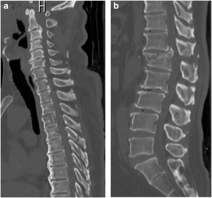
We report the successful conservative management of an unusual case of esophageal perforation associated with an upper thoracic spinal fracture from blunt trauma in Minamata, Kumamoto, Japan. A 69-year-old man became paraplegic secondary to an L1 burst fracture caused by a boating accident and underwent posterior fixation on the day of admission. The patient also had a minimally displaced T4 vertebral fracture. Fever, dyspnea and elevated inflammatory markers all persisted postoperatively. Computed tomography showed free mediastinal air at the T4 level, and an esophagram showed contrast medium leakage, which helped diagnose esophageal perforation. The esophageal perforation healed with conservative treatment without life-threatening complications. The possibility of esophageal injury should always be considered when treating upper thoracic spinal injuries due to blunt trauma.
Keywords: Neurosurgery; Trauma.
Figures

References
-
- Beal SL , Pottmeyer EW , Spisso JM . Esophageal perforation following external blunt trauma. J Trauma 1988; 28: 1425–1432. - PubMed
-
- Jones WG II , Ginsberg RJ . Esophageal perforation: a continuing challenge. Ann Thorac Surg 1992; 53: 534–543. - PubMed
-
- Chen HC , Tzaan WC , Chen TY , Tu PH . Esophageal perforation complication with spinal epidural abscess, iatrogenic or secondary to first thoracic spine fracture? Acta Neurochir (Wien) 2005; 147: 431–434. - PubMed
-
- Chen SH , Huang TJ , Chen YJ , Liu HP , Hsu RW . Flexion-dislocation injury of the upper thoracic spine associated with tracheoesophageal perforation: a case report. J Bone Joint Surg Am 2002; 84: 1028–1031. - PubMed
LinkOut - more resources
Full Text Sources
Other Literature Sources
