. 2016 Sep 15:2:16020.
doi: 10.1038/scsandc.2016.20. eCollection 2016.

Diagnostic challenges in vacuolar myelopathy: a didactic case report

Affiliations

Diagnostic challenges in vacuolar myelopathy: a didactic case report

Florian Ernst et al. Spinal Cord Ser Cases. .

Abstract

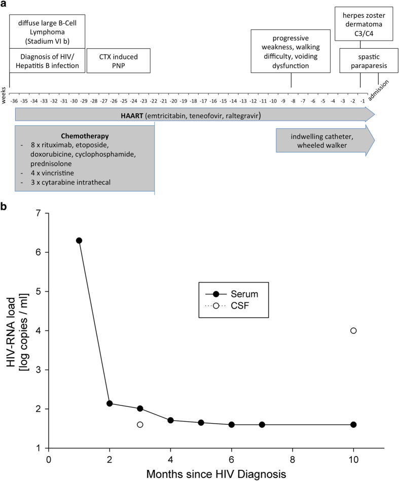
Introduction: Because of the diagnostic complexity and potential pitfalls in interpreting test results, HIV-vacuolar myelopathy (HIVM) is far more often diagnosed postmortem than in vivo. In the era of highly active antiretroviral therapy (HAART), the topic of neuro-AIDS has become increasingly important. This case report covers some of the diagnostic problems encountered in vacuolar myelopathy based on magnetic resonance imaging (MRI) fiber-tracking pictures of the spine in a patient with HIVM, including a 1-year follow-up.

Case presentation: A 49-year-old man felt progressive weakness, and difficulties while walking, and he suffered from incomplete voiding. A week before admission, follicles appeared on the right side of his neck and shoulder. His medical history included a chronic HIV infection treated with HAART and a B-cell lymphoma in complete remission after chemotherapy. The initial exam revealed thoracic hyposensitivity level distal to dermatome Th9, spastic paraparesis of the lower limbs and herpes zoster infection in dermatome C3/C4. A lesion of the thoracic myelon could be ruled out in the MRI scan, chemotherapy-induced polyneuropathy was stable, and no acute opportunistic infection of the CNS was found. HIV load in cerebrospinal fluid (CSF) was markedly elevated. An HIV-associated vacuolar myelopathy was diagnosed, revealing the HIV itself as etiology.

Discussion: A negative or unspecific MRI scan excludes possible other causes, but by no means rules out HIV-related myelopathy. Furthermore, peripheral and central viral load should always be assessed to avoid missing a possible 'CSF HIV-escape'.

Keywords: Central nervous system infections; Disability; Infection; Neurological manifestations; Spinal cord.


Figures

Figure 1
Figure 1
(a, b) Course of the disease with serum and CSF HIV-RNA load. (a) The timeline of diagnoses, therapies and symptoms. The patient underwent regular checks in the outpatient clinic for infectious diseases, where HIV-RNA load was measured in serum. After the start of HAART, the viral load dropped significantly below the detection limit. The CSF viral load was measured once at the time point of HIV diagnosis and the second time when the patient was admitted to our department. A clear heightened viral load in CSF can be seen as a possible ‘CSF HIV-escape’ (b).
Figure 2
Figure 2
(ad) Magnetic resonance images of the head and thoracic spine. Cerebral MRI (sagittal T1 weighted (a), coronal (b) and transversal (c) post-gadolinium T1-weighted images) merely shows discrete signs of leukoencephalopathy but no contrast agent enhancement, whereas spinal MRI (sagittal T2-weighted images of the thoracic spine (d)) reveals a reduced bone marrow signal without myelopathy signal and without contrast agent enhancement. The cervical and thoracic cords appear atrophic with an anteroposterior diameter of 5–6 and 4–5 mm, respectively. On the sagittal T2 images, no definitive hyperintense areas are visible.
Figure 3
Figure 3
(ae) Magnetic resonance images and tractography of the whole spine (data collection within the follow-up one and a half years later). As an overview, sagittal post-gadolinium T1-weighted spectral presaturation with inversion recovery (SPIR) sequences of the whole spine were performed (a). The diffusion tensor imaging and tractography study was performed on a whole-body 3.0-T scanner (Achieva, Philips Medical Systems, Best, Netherlands). Diffusion tensor (DT) images were obtained using a DwiSE scan technique with the following parameters: TR/TE=3823.4 ms/83.3 ms; flip angle 90°; field of view (FOV) 224 mm; number of signals averaged (NSA), two; 32-noncollinear diffusion gradient directions; b-values, 0 and 800 s mm−2; isotropic voxel size of 2 mm; SENSE technique; spine coil. Sixty slices of DT images on the axial plane were acquired from the mid of the Th7 spine level to the inferior line of Th10. Post-processing analysis was performed on a Philips Extended MR Workspace R2.6.3.1 (Philips Medical Systems, Hamburg, Germany). The Philips FiberTrack package permits streamline tractography with single and multiple regions of interest (ROIs). Algorithm settings are as follows: minimum FA 0.15; maximum angle change (deg) 27.0; and minimum fiber length 10 mm. We performed streamline (‘deterministic’) tractography with single and multiple regions of interest in the spinal cord. This technique is based on the FACT (fiber assignment by continuous tracking) algorithm, which, in short, connects the largest eigenvectors (with the largest eigenvalue) of the diffusion ellipsoid of the voxels of the spinal cord according to the algorithm setting. The results are streamlines, which depict the main diffusion direction (‘axonal direction’). We did not measure the two minor eigenvalues of the diffusion ellipsoid; therefore, no specific information about ‘myelination’ is available. Three-dimensional single ROI tractography in the anterior and lateral view from thoracic level 7 to 10. At thoracic vertebral level 7, one voxel could be placed into the ADC-map of the spinal cord with an FA value of 0.724 and an ADC of 0.744×10⁻³ mm2/s (b). At thoracic vertebral level 8, one voxel could be placed somewhat excentrically into the spinal cord with an FA value of 0.764 and an ADC of 0.741×10⁻³ mm2 s−1 (c). The lower portion of the spinal cord appeared too small and distorted to place a voxel centrally. Geometric distortion did not allow an exact superposition of the axially reconstructed FA maps on anatomical images. To summarize, the diffusion tensor imaging shows many streamlines in the longitudinal direction of the spinal cord, connecting anisotropic voxels, and did not reveal severe disruptions, but was somewhat degraded by step artifacts due to susceptibility effects, motion and geometric distortion (d, e).

References

    1. Dal Pan GJ, Glass JD, McArthur JC. Clinicopathologic correlations of HIV-1-associated vacuolar myelopathy: an autopsy-based case-control study. Neurology 1994; 44: 2159–2164. - PubMed
    1. Chong J, Di Rocco A, Tagliati M, Danisi F, Simpson DM, Atlas SW. MR findings in AIDS-associated myelopathy. AJNR Am J Neuroradiol 1999; 20: 1412–1416. - PMC - PubMed
    1. Kwong Y-L, Yeung DYM, Chan JCW. Intrathecal chemotherapy for hematologic malignancies: drugs and toxicities. Ann Hematol 2008; 88: 193–201. - PubMed
    1. Cho TA, Vaitkevicius H. Infectious myelopathies. Continuum (Minneapolis Minn.) 2012; 18: 1351–1373. - PubMed
    1. Bizaare M, Dawood H, Moodley A. Vacuolar myelopathy: a case report of functional, clinical, and radiological improvement after highly active antiretroviral therapy. Int J Infect Dis 2008; 12: 442–444. - PubMed

LinkOut - more resources