Review
. 2016 Dec 18:5:5-14.
doi: 10.1016/j.dadm.2016.12.006. eCollection 2016.

Imaging markers of cerebrovascular pathologies: Pathophysiology, clinical presentation, and risk factors

Affiliations
Review

Imaging markers of cerebrovascular pathologies: Pathophysiology, clinical presentation, and risk factors

Mekala R Raman et al. Alzheimers Dement (Amst). .

Abstract

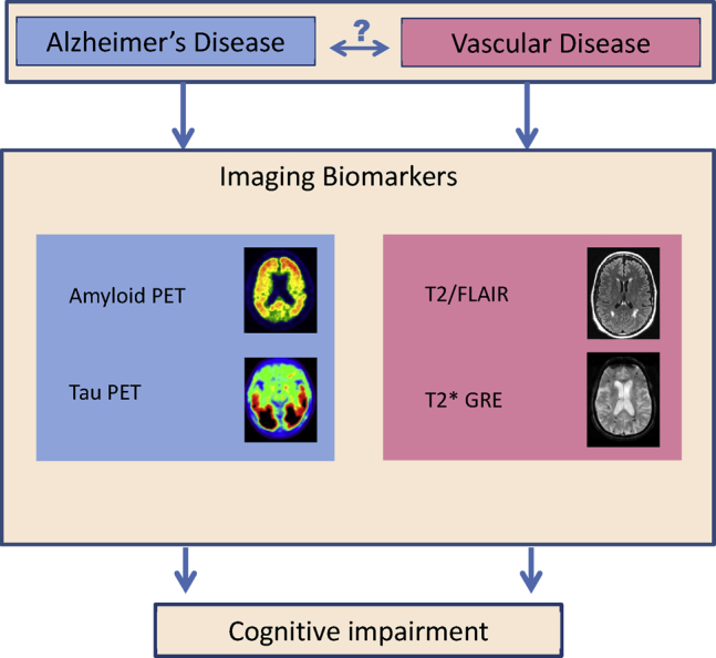
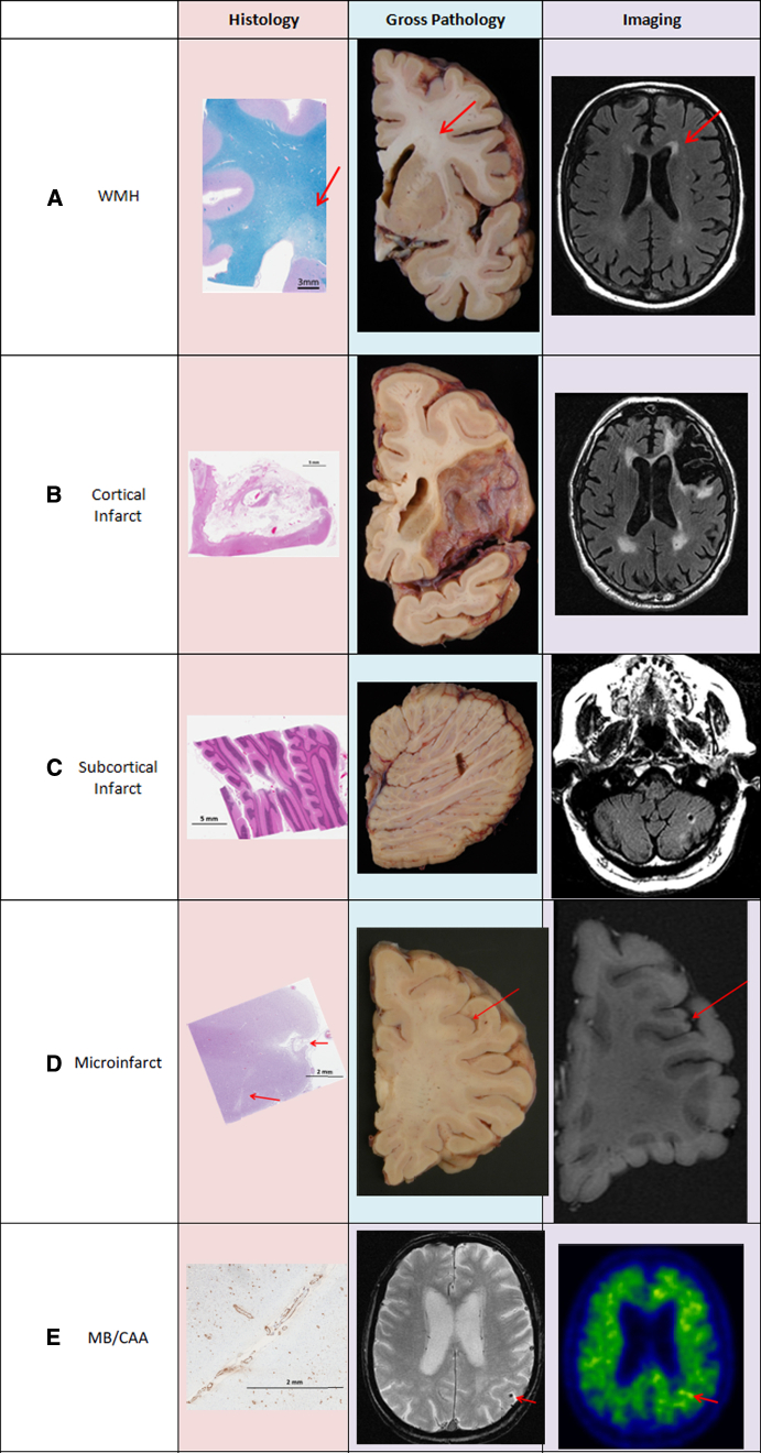
Cerebrovascular pathologies (CVPs) are common pathologies associated with age-related cognitive decline along with Alzheimer disease pathologies. The impact of CVP on the prevalence of dementia is increasingly being recognized. The goal of this review is to improve our understanding of the pathophysiological underpinnings and the multimodal magnetic resonance imaging and positron emission tomography imaging changes that are associated with the hallmarks of CVP. This knowledge will facilitate the development of early detection, intervention, and prevention strategies that may contribute to lowering the risk of dementia. In this review, we will first discuss currently known risk factors of CVPs including cardiovascular, lifestyle, genetic, sex differences, and head injury. Next, we will focus on the pathophysiology of CVPs and their impact on neurodegeneration and downstream cognitive impairment. Specifically, we will discuss three of the most common cerebrovascular lesions seen on MRI: white-matter hyperintensity, microbleeds, and infarcts. Finally, we will discuss the unanswered open questions in this field.

Keywords: Aging; Cerebrovascular; Imaging; Pathophysiology.


Figures

Fig. 1
Fig. 1
Alzheimer disease has positron emission tomorgraphy imaging biomarkers (left) that are available for assessing the accumulation of amyloid β and tau pathologies. Only surrogates of cerebrovascular pathology using imaging are possible. T2/FLAIR and T2* GRE magnetic resonancy imaging (right) provide surrogates of ischemic pathology and microhemorrhages, respectively. The relationship between Alzheimer disease and vascular disease in their contributions to cognitive impairment is still unknown. Abbreviations: FLAIR, Fluid Attenuated Inversion Recovery; GRE, gradient recalled echo.
Fig. 2
Fig. 2
Cerebrovascular disease pathologies on histology, gross pathology, and imaging. (A) White-matter hyperintensities: rarefied tissue (indicated by arrows) on histology, discolored white matter on gross pathology, and hyperintensity on FLAIR MRI. (B) Large cortical infarct: tissue loss and gliosis on H&E stained histology slide, apparent tissue loss on gross pathology, tissue loss with surrounding hyperintensity. (C) Subcortical cerebellar infarct: tissue loss and gliosis on H&E stained histology slide, apparent tissue loss on gross pathology, tissue loss with surrounding hyperintensity. (D) Cortical microinfarct (indicated by arrows): tissue loss and gliosis on H&E stained histology slide, not apparent on gross pathology, microinfarcts >1 mm seen on high-resolution MRI. (E) Lobar microbleeds/CAA: amyloid uptake in vessel walls seen on parietal lobe histology slide with amyloid stain, not apparent on gross pathology, microbleed visible in parietal lobe (shown by arrow), and increased focal uptake seen on amyloid PET scan (shown by arrow). Abbreviations: CAA, cerebral amyloid angiopathy; FLAIR, Fluid Attenuated Inversion Recovery; GRE, gradient recalled echo; WMH, white-matter hyperintensity.

References

    1. Schneider J.A., Aggarwal N.T., Barnes L., Boyle P., Bennett D.A. The neuropathology of older persons with and without dementia from community versus clinic cohorts. J Alzheimers Dis. 2009;18:691–701. - PMC - PubMed
    1. Jellinger K.A. Understanding the pathology of vascular cognitive impairment. J Neurol Sci. 2005;229-230:57–63. - PubMed
    1. Nelson P.T., Head E., Schmitt F.A., Davis P.R., Neltner J.H., Jicha G.A. Alzheimer's disease is not “brain aging”: neuropathological, genetic, and epidemiological human studies. Acta Neuropathol. 2011;121:571–587. - PMC - PubMed
    1. Jellinger K.A. Pathology and pathogenesis of vascular cognitive impairment-a critical update. Front Aging Neurosci. 2013;5:17. - PMC - PubMed
    1. Vemuri P., Knopman D.S. The role of cerebrovascular disease when there is concomitant Alzheimer disease. Biochim Biophys Acta. 2016;1862:952–956. - PMC - PubMed