. 2017 Jan 18;139(2):863-870.
doi: 10.1021/jacs.6b11082. Epub 2017 Jan 5.

Guanidinium can both Cause and Prevent the Hydrophobic Collapse of Biomacromolecules

Affiliations

Guanidinium can both Cause and Prevent the Hydrophobic Collapse of Biomacromolecules

Jan Heyda et al. J Am Chem Soc. .

Abstract

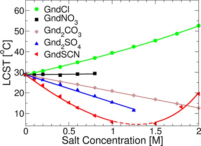
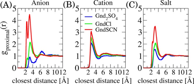
A combination of Fourier transform infrared and phase transition measurements as well as molecular computer simulations, and thermodynamic modeling were performed to probe the mechanisms by which guanidinium (Gnd+) salts influence the stability of the collapsed versus uncollapsed state of an elastin-like polypeptide (ELP), an uncharged thermoresponsive polymer. We found that the cation's action was highly dependent upon the counteranion with which it was paired. Specifically, Gnd+ was depleted from the ELP/water interface and was found to stabilize the collapsed state of the macromolecule when paired with well-hydrated anions such as SO42-. Stabilization in this case occurred via an excluded volume (or depletion) effect, whereby SO42- was strongly partitioned away from the ELP/water interface. Intriguingly, at low salt concentrations, Gnd+ was also found to stabilize the collapsed state of the ELP when paired with SCN-, which is a strong binder for the ELP. In this case, the anion and cation were both found to be enriched in the collapsed state of the polymer. The collapsed state was favored because the Gnd+ cross-linked the polymer chains together. Moreover, the anion helped partition Gnd+ to the polymer surface. At higher salt concentrations (>1.5 M), GndSCN switched to stabilizing the uncollapsed state because a sufficient amount of Gnd+ and SCN- partitioned to the polymer surface to prevent cross-linking from occurring. Finally, in a third case, it was found that salts which interacted in an intermediate fashion with the polymer (e.g., GndCl) favored the uncollapsed conformation at all salt concentrations. These results provide a detailed, molecular-level, mechanistic picture of how Gnd+ influences the stability of polypeptides in three distinct physical regimes by varying the anion. It also helps explain the circumstances under which guanidinium salts can act as powerful and versatile protein denaturants.


Conflict of interest statement

The authors declare no competing financial interest.

Figures

Figure 1
Figure 1
(A) Schematic illustration of how Gnd2SO4 makes the collapsed state of the polypeptide favorable via ion exclusion. (B) Schematic illustration of how GndSCN makes the collapsed state of polypeptide more favorable (at <1 M salt concentration) via guanidinium inclusion along with thiocyanate binding to the backbone as illustrated in the zoomed-in picture. This behavior switches over to favoring the uncollapsed polypeptide upon additional ion inclusion at higher salt concentration (>1.5 M).
Figure 2
Figure 2
LCST measurements of 10 mg/mL ELP solutions as a function of guanidinium salt concentration. All standard deviations were within the data points drawn. Each symbol represents data points from six measurements, and the solid lines are fits to eq 2. No data were obtained between 1.0 and 1.5 M GdnSCN, where the LCST value fell below 4 °C (dashed portion of the red line).
Figure 3
Figure 3
(A) Experimental scheme showing an aqueous solution droplet (100 μL) placed onto the diamond coated ZnSe ATR crystal. The upper left insert represents an initially homogeneous polypeptide solution (below the LCST), whereas the upper right inset, represents the ATPS which is formed above the LCST. (B) Fitted ATR-FTIR spectra of the collapsed state of the ELP above the LCST (45 °C) in the presence of 0.5 M d-GndSCN, in D2O in the amide I spectral region, and (C) in the C=N stretch band region of SCN. (D) Fitted spectra of the same solution except in the presence of 0.5 M d-Gnd2SO4 in D2O in the amide I spectral region, along with (E) the vibrational spectra of the S—O stretching band of SO42–. (F) Plots of the amide I spectral region with and without the macromolecule in solutions containing 0.5 M d-GndCl. In panels B–F, the red and blue curves indicate data taken in the presence of the ELP and in its absence, respectively. The gray lines represent three Gaussian fits to the amide I bands, whereas the green curves represent the overall measured spectra. The inset schematics in panels C, E, and F depict ion accumulation for GndSCN and ion depletion for Gnd2SO4 along with only slight ion accumulation for GndCl. The asterisks in panel E denote weak fingerprint vibrational resonances related to polypeptide.
Figure 4
Figure 4
(A) Results of coarse-grained simulations for a model polymer in cosolvent solutions that bind strongly (red), weakly (green), or are depleted from the polymer surface (blue). The results plot the radius of gyration of the polymer (scaled by that of an ideal chain with the same bond length) as a function of cosolvent concentration. (B) Distribution of cosolvent from the center of the polymer at 1 M concentration. The inset presents the same distribution curves at 13 M cosolvent. In panel B, r denotes the distance to center of mass of the polymer. (C) Schematic depiction of the mechanism of swelling and compression of the polymer (red spheres) caused by the cosolvent moieties (yellow spheres) in the low (1 M) and high (13 M) concentration regimes. Note that the existence of multivalent binding interactions of the cosolvent at low solvent concentration is responsible for the polymer collapse in the strong binding regime. Only the cosolvent molecules in direct contact (<4 Å) with the polymer chain are depicted for clarity.
Figure 5
Figure 5
Spatial density maps of the ions, as obtained from all atom MD simulations. (A) This snapshot shows the depletion of Gnd2SO4 (Gnd+ in purple and SO42– in silver) from the vicinity of the VPGVG pentapeptide. (B) GndCl (Cl in orange) and (C) GndSCN (SCN in yellow). The contours plotted for each ion correspond to 4× the bulk density for all three snapshots.
Figure 6
Figure 6
Proximal distribution function of the investigated guanidinium salts around the extended VPGVG pentapeptide. The distribution of anions is shown in the panel A, that of the Gnd+ cation is in the panel B, and overall salt distribution in the panel C.

References

    1. Jungwirth P.; Cremer P. S. Beyond Hofmeister. Nat. Chem. 2014, 6, 261–263. 10.1038/nchem.1899. - DOI - PubMed
    1. Kunz W.; Lo Nostro P.; Ninham B. W. The Present State of Affairs with Hofmeister Effects. Curr. Opin. Colloid Interface Sci. 2004, 9, 1–18. 10.1016/j.cocis.2004.05.004. - DOI
    1. Leontidis E. Hofmeister Anion Effects on Surfactant Self-Assembly and the Formation of Mesoporous Solids. Curr. Opin. Colloid Interface Sci. 2002, 7, 81–91. 10.1016/S1359-0294(02)00010-9. - DOI
    1. Zhang Y.; Cremer P. S. The Inverse and Direct Hofmeister Series for Lysozyme. Proc. Natl. Acad. Sci. U. S. A. 2009, 106, 15249–15253. 10.1073/pnas.0907616106. - DOI - PMC - PubMed
    1. Paterova J.; Rembert K. B.; Heyda J.; Kurra Y.; Okur H. I.; Liu W. S. R.; Hilty C.; Cremer P. S.; Jungwirth P. Reversal of the Hofmeister Series: Specific Ion Effects on Peptides. J. Phys. Chem. B 2013, 117, 8150–8158. 10.1021/jp405683s. - DOI - PubMed

Publication types

LinkOut - more resources