. 2017 Jan 5:7:39899.
doi: 10.1038/srep39899.

Hepatic MiR-291b-3p Mediated Glucose Metabolism by Directly Targeting p65 to Upregulate PTEN Expression

Affiliations

Hepatic MiR-291b-3p Mediated Glucose Metabolism by Directly Targeting p65 to Upregulate PTEN Expression

Jun Guo et al. Sci Rep. .

Abstract

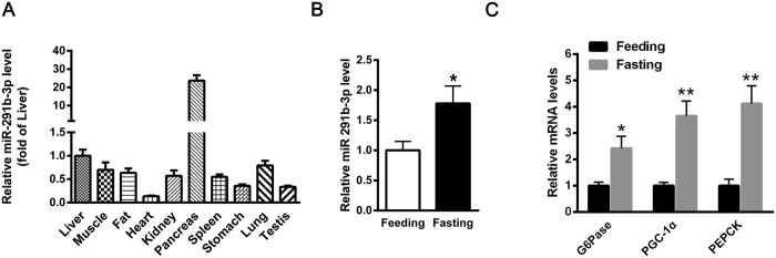
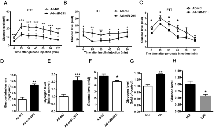
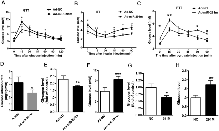
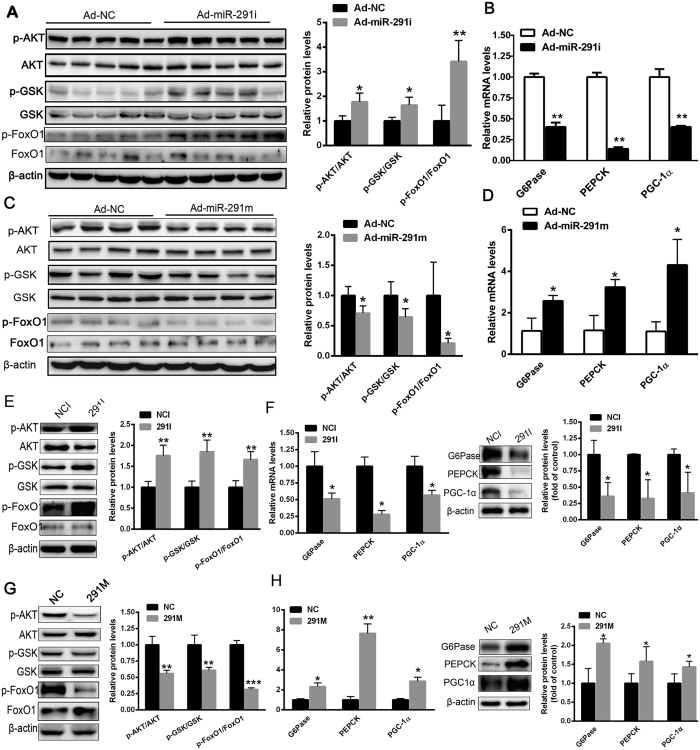
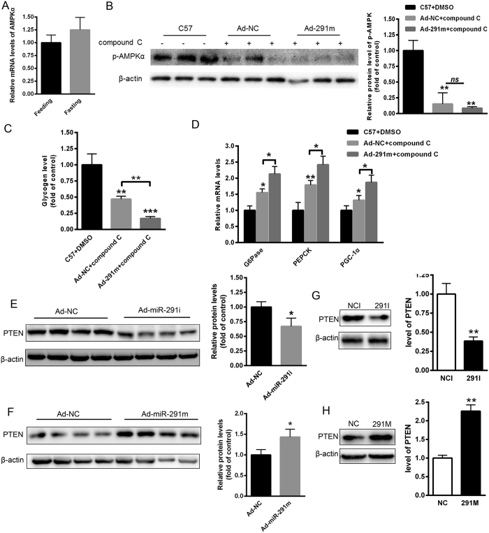
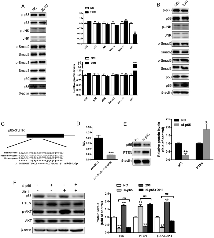
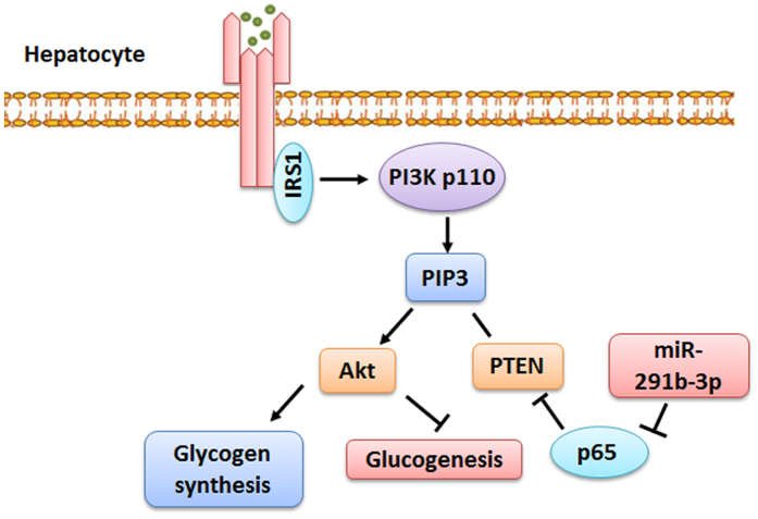
Several studies have suggested an important role of miR-291b-3p in the development of embryonic stem cells. In previous study, we found that the expression of miR-291b-3p was significantly upregulated in the liver of db/db mice. However, the role of miR-291b-3p in glucose metabolism and its underlying mechanisms remain unknown. In the present study, we demonstrated that miR-291b-3p was abundantly expressed in the liver. Of note, hepatic miR-291b-3p expression was upregulated in HFD-fed mice and induced by fasting in C57BL/6 J normal mice. Importantly, hepatic inhibition miR-291b-3p expression ameliorated hyperglycemia and insulin resistance in HFD-fed mice, whereas hepatic overexpression of miR-291b-3p led to hyperglycemia and insulin resistance in C57BL/6 J normal mice. Further study revealed that miR-291b-3p suppressed insulin-stimulated AKT/GSK signaling and increased the expression of gluconeogenic genes in hepatocytes. Moreover, we identified that p65, a subunit of nuclear factor-κB (NF-κB), is a target of miR-291b-3p by bioinformatics analysis and luciferase reporter assay. Silencing of p65 significantly augmented the expression of PTEN and impaired AKT activation. In conclusion, we found novel evidence suggesting that hepatic miR-291b-3p mediated glycogen synthesis and gluconeogenesis through targeting p65 to regulate PTEN expression. Our findings indicate the therapeutic potential of miR-291b-3p inhibitor in hyperglycemia and insulin resistance.


Figures

Figure 1
Figure 1. Hepatic miR-291b-3p expression is upregulated in HFD-fed mice and induced by fasting in C57BL/6 J normal mice.
(A) The expression of miR-291b-3p in different organs of C57BL/6 J normal mice, as analyzed by real-time PCR. (B) The expression of miR-291b-3p in the liver of C57BL/6 J mice fasted for 16 h. (C) The mRNA levels of G6Pase, PGC1-α and PEPCK were determined in the liver of fasted mice. Data are expressed as mean ± SEM (n = 5). *P < 0.05; **P < 0.01, ***P < 0.001 vs. other group.
Figure 2
Figure 2. Hepatic inhibition miR-291b-3p expression ameliorates hyperglycemia and insulin resistance in HFD-fed mice.
(A) Glucose tolerance test (GTT) in HFD-fed mice on 7 days after injection of adenovirus-miR-291b-3p-inbibitor (Ad-miR-291i) or adenovirus-GFP (Ad-NC). (B) Insulin tolerance test (ITT) in HFD-fed mice on 7 days after injection of Ad-miR-291i or Ad-NC. (C) Pyruvate tolerance test (PTT) in HFD-fed mice on 7 days after injection of Ad-miR-291i or Ad-NC. (D) Euglycemic-hyperinsulinemic clamp assay in HFD-fed mice on 7 days after injection of Ad-miR-291i or Ad-NC. (E) The glycogen content in the liver of HFD-fed mice on 7 days after injection of Ad-miR-291i or Ad-NC. (F) The glucose production in the liver of HFD-fed mice on 7 days after injection of Ad-miR-291i or Ad-NC. (G) The glycogen content in the NCTC1469 cells transfected with miR-291b-3p inhibitor (291i). (H) The glucose level in the medium of NCTC1469 cells transfected with 291i. Data are expressed as mean ± SEM (n = 5). *P < 0.05; **P < 0.01 vs. control group.
Figure 3
Figure 3. Hepatic overexpression of miR-291b-3p results in hyperglycemia and insulin resistance in C57BL/6 J mice.
(A) GTT in C57BL/6 J mice on 7 days after injection of adenovirus- miR-291b-3p-mimic (Ad-miR-291m) or adenovirus-GFP (Ad-NC). (B) ITT in C57BL/6 J mice on 7 days after injection of Ad-miR-291m or Ad-NC. (C) PTT in C57BL/6 J mice on 7 days after injection of Ad-miR-291m or Ad-NC. (D) Euglycemic-hyperinsulinemic clamp assay in C57 mice on 7 days after injection of Ad-miR-291m or Ad-NC. (E) The glycogen content in the liver of C57BL/6 J mice on 7 days after injection of Ad-miR-291m or Ad-NC. (F) The glucose production in the liver of C57BL/6 J mice on 7 days after injection of Ad-miR-291m or Ad-NC. (G) The glycogen content in the NCTC1469 cells transfected with miR-291b-3p mimic (miR-291m). (H) The glucose level in the medium of NCTC1469 cells transfected with miR-291m. Data are expressed as mean ± SEM (n = 5). *P < 0.05; **P < 0.01; ***P < 0.001 vs. control group.
Figure 4
Figure 4. MiR-291b-3p suppresses insulin-stimulated AKT/GSK/FoxO1 signaling and increases expression of gluconeogenic genes in hepatocytes.
(A) The phosphorylation of AKT/GSK/FoxO1 in the liver of HFD-fed mice on 7 days after injection of Ad-miR-291i or Ad-NC. Full-length blots/gels are presented in Supplementary Figure 1. (B) The mRNA levels of gluconeogenesis genes including G6Pase, PGC-1α and PEPCK in the liver of HFD-fed mice on 7 days after injection of Ad-miR-291i or Ad-NC. (C) The phosphorylation of AKT/GSK/FoxO1 in the liver of C57BL/6 J mice on 7 days after injection of Ad-miR-291m or Ad-NC. Full-length blots/gels are presented in Supplementary Figure 2 and cropping lines are indicated in red color. (D) The mRNA levels of G6Pase, PGC-1α and PEPCK in the liver of C57BL/6 J mice on 7 days after injection of Ad-miR-291m or Ad-NC. (E) The phosphorylation of AKT/GSK/FoxO1 in the NCTC1469 cells transfected with miR-291b-3p inhibitor (291i). Full-length blots/gels are presented in Supplementary Figure 3 and cropping lines are indicated in red color. (F) The mRNA and protein levels of gluconeogenesis genes in the NCTC1469 cells transfected with 291i. (G) The phosphorylation of AKT/GSK/FoxO1 in the NCTC1469 cells transfected with miR-291b-3p mimic (291 m). Full-length blots/gels are presented in Supplementary Figure 4 and cropping lines are indicated in red color. (H) The mRNA and protein levels of G6Pase, PGC-1α and PEPCK in the NCTC1469 cells transfected with 291 m. Data are expressed as mean ± SEM (n = 5). *P < 0.05; **P < 0.01 vs. control group.
Figure 5
Figure 5. MiR-291b-3p elevates the expression of PTEN.
(A) The mRNA level of AMPK in the liver of fasted mice. (B) The hepatic phosphorylation level of AMPK in the C57 mice injected with compound c, an AMPK inhibitor, at a dose of 2 mg/kg for 3 days. (C) The glycogen level in the liver of C57 mice transfected with Ad-NC and Ad-291m, and injected with compound C. (D) The mRNA levels of G6Pase, PGC1-α and PEPCK in the liver of C57 mice transfected with Ad-NC and Ad-291m, and injected with compound C. (E) The hepatic level of PTEN protein in the liver of HFD-fed mice on 7 days after injection of Ad-miR-291i or Ad-NC. (F) The hepatic expression of PTEN protein in the liver of C57BL/6 J mice on 7 days after injection of Ad-miR-291i or Ad-NC. (G) The protein level of PTEN in the NCTC1469 cells transfected with 291i or NC. (H) The expression of PTEN protein in the NCTC1469 cells transfected with 291 m or NC. Full-length blots/gels are presented in Supplementary Figure 5 and cropping lines are indicated in red color. Data are expressed as mean ± SEM (n = 5). *P < 0.05; **P < 0.01 vs. control group.
Figure 6
Figure 6. MiR-291b-3p directly targets p65 to upregulate PTEN expression.
(A) The expression or activation of transcription factors and pathways regulating PTEN expression, including p38MAPK, JNK, Smad2/3 and NF-κB (p50 and p65) in the NCTC1469 cells transfected with 291 m or NC. Full-length blots/gels are presented in Supplementary Figure 6 and cropping lines are indicated in red color (left pannel). (B) The expression or activation of p38MAPK, JNK, Smad2/3 and NF-κB (p50 and p65) in the NCTC1469 cells transfected with 291i or NC. Full-length blots/gels are presented in Supplementary Figure 6 and cropping lines are indicated in red color (right pannel). (C) A conserved binding site of the 3′UTR of p65 with miR-291b-3p. (D) Relative luciferase activity in the NCTC1469 cells transfected with reporter constructs containing the 3′UTR of mouse p65 gene. (E) The levels of p65 and PTEN protein in the NCTC1469 cells transfected with a specific siRNA targeting p65. Full-length blots/gels are presented in Supplementary Figure 7A. (F) The levels of p65, PTEN, AKT and AKT phosphorylation in the NCTC1469 cells transfected with p65, miR-291b-3p inhibitor alone or both. Full-length blots/gels are presented in Supplementary Figure 7B and cropping lines are indicated in red color. Data are expressed as mean ± SEM (n = 3 independent experiments). *P < 0.05; **P < 0.01 vs. control group.
Figure 7
Figure 7. Proposed mechanisms by which hepatic miR-291b-3p regulates insulin signaling and glucose metabolism.
MiR-291b-3p targets p65 to regulate PTEN expression, in turn mediated glycogen synthesis and gluconeogenesis in hepatocytes.

Similar articles

Cited by

References

    1. Abu-Farha M. et al.. Higher plasma betatrophin/ANGPTL8 level in Type 2 Diabetes subjects does not correlate with blood glucose or insulin resistance. Scientific reports 5, 10949 (2015). - PMC - PubMed
    1. Tangvarasittichai S. Oxidative stress, insulin resistance, dyslipidemia and type 2 diabetes mellitus. World journal of diabetes 6, 456–480 (2015). - PMC - PubMed
    1. Nolan C. J., Ruderman N. B., Kahn S. E., Pedersen O. & Prentki M. Insulin resistance as a physiological defense against metabolic stress: implications for the management of subsets of type 2 diabetes. Diabetes 64, 673–686 (2015). - PMC - PubMed
    1. Chakraborty C., Doss C. G., Bandyopadhyay S. & Agoramoorthy G. Influence of miRNA in insulin signaling pathway and insulin resistance: micro-molecules with a major role in type-2 diabetes. Wiley interdisciplinary reviews. RNA 5, 697–712 (2014). - PubMed
    1. Chang C. C. et al.. Interferon gamma-induced protein 10 is associated with insulin resistance and incident diabetes in patients with nonalcoholic fatty liver disease. Scientific reports 5, 10096 (2015). - PMC - PubMed

Publication types