. 2017 Jan 5:7:39975.
doi: 10.1038/srep39975.

High Shear Stresses under Exercise Condition Destroy Circulating Tumor Cells in a Microfluidic System

Affiliations

High Shear Stresses under Exercise Condition Destroy Circulating Tumor Cells in a Microfluidic System

Sagar Regmi et al. Sci Rep. .

Abstract

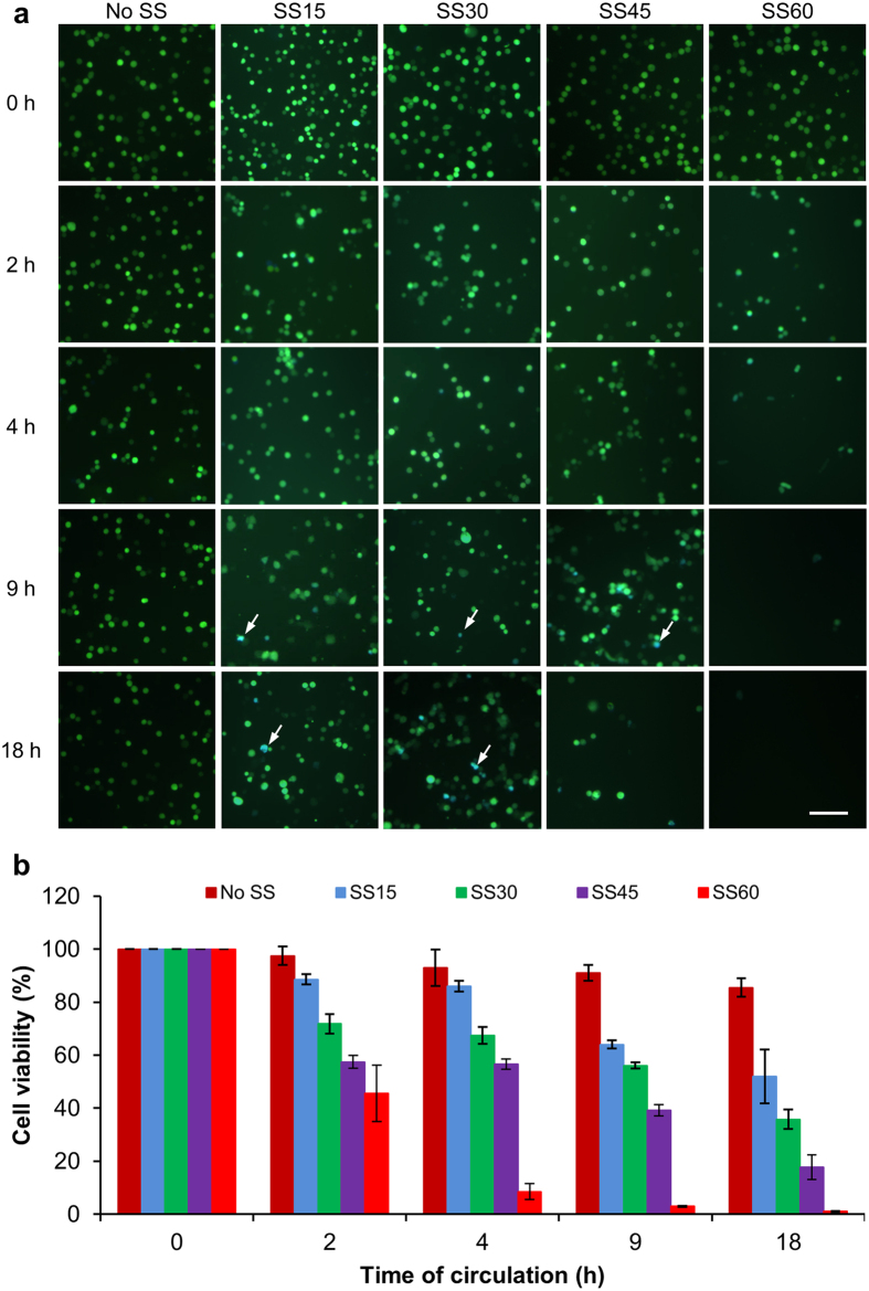
Circulating tumor cells (CTCs) are the primary targets of cancer treatment as they cause distal metastasis. However, how CTCs response to exercise-induced high shear stress is largely unknown. To study the effects of hemodynamic microenvironment on CTCs, we designed a microfluidic circulatory system that produces exercise relevant shear stresses. We explore the effects of shear stresses on breast cancer cells with different metastatic abilities, cancer cells of ovarian, lung and leukemic origin. Three major findings were obtained. 1) High shear stress of 60 dynes/cm2 achievable during intensive exercise killed more CTCs than low shear stress of 15 dynes/cm2 present in human arteries at the resting state. 2) High shear stress caused necrosis in over 90% of CTCs within the first 4 h of circulation. More importantly, the CTCs that survived the first 4 h-circulation, underwent apoptosis during 16-24 h of post-circulation incubation. 3) Prolonged high shear stress treatment effectively reduced the viability of highly metastatic and drug resistant breast cancer cells. As high shear stress had much less damaging effects on leukemic cells mimicking the white blood cells, we propose that intensive exercise may be a good strategy for generating high shear stress that can destroy CTCs and prevent cancer metastasis.


Figures

Figure 1
Figure 1. Design of the microfluidic circulatory system.
(a) Schematic diagram of the microfluidic circulatory system that can generate various levels of SS. Correlations between the speed settings of the peristaltic pump with the flow rate (b) and levels of SS (c).
Figure 2
Figure 2. Shear force- and time-dependent death of 231-C3 cells.
(a) Merged FRET images of 231-C3 cells immediately after being treated with No SS, SS15, SS30, SS45 and SS60 for 0, 2, 4, 9 and 18 h. The white arrows indicate the blue apoptotic cells. Scale bar: 100 μm. (b) Viability of 231-C3 cells was measured by the MTT assay under the same conditions indicated in panel (a). The results were normalized to 0 h.
Figure 3
Figure 3. Examination of the SS effects.
(a) Phase and merged FRET images of the 231-C3 cells after 0, 2, 3 and 4 h of circulation under the treatment of SS15 and SS60. Scale bar: 100 μm. (b) Viability of 231-C3 cells was measured by the MTT assay under the same treatment. The results were normalized to 0 h. ***p < 0.001.
Figure 4
Figure 4. Examination of the necrotic effects of high SS on circulated 231-C3 cells.
(a) Optical micrograph of 231-C3 cells that were circulated under SS15 and SS60 for 4 h. Necrotic cells were visualized using propidium iodide (PI) staining, while apoptotic cells were indicated by FRET imaging. Necrotic cells emit a red color, whereas the live cells emit a green color. The white arrows indicate the red necrotic cells. Scale bar: 100 μm. (b) Relation of LDH release with the time (1–4 h) of SS treatment. The results were normalized to 0 h.
Figure 5
Figure 5. Cell fate after exposure to low and high SS.
The cells were exposed to SS15 and SS60 for 4 h and allowed to recover under a static condition. The images were captured during the post-circulation period at the indicated times. (a) Merged FRET images of the 231-C3 cells at 12, 16 and 24 h post-circulation under SS15 and SS60 for 4 h. The white arrows indicate the blue apoptotic cells. Scale bar: 100 μm. (b) The viability of the 231-C3 cells was measured by the MTT assay under the same treatment conditions as in (a). The results were normalized to before circulation. (c) The percentage of apoptotic cells was quantified from the FRET images (n = 300 cells, ***p < 0.001).
Figure 6
Figure 6. Examining the cytotoxic effects of SS on multiple types of cancer cells.
(a) Optical micrographs showing the effects of the low and high SS conditions on three cell lines of A549, 2008 and UACC-893. (b) The cell viability was measured by the MTT assay after 4 h of circulation under SS15 and SS60. ***p < 0.001.
Figure 7
Figure 7. Effects of high SS treatments on cancer cells with different metastatic and drug resistant abilities.
(a) Merged FRET images of 231-C3, 231-M1 and 231-M1A cells after 4 and 8 h of SS60 treatment. Scale bar: 100 μm. (b) Viability of 231-series cells was measured by the MTT assay at 4 and 8 h after SS60 treatment. The results were normalized to 0 h. ***p < 0.001 vs. 231-C3 cells.
Figure 8
Figure 8. Leukemic cells are highly resistant against strong shear forces.
(a) Optical micrographs showing the effects of the low and high SS conditions on K562 cells. Scale bar: 200 μm. (b) The viability of K562 cells was measured by the MTT assay after 1–4 h of SS15 and SS60 treatment. The results were normalized to 0 h. *p < 0.05, **p < 0.01.

Similar articles

Cited by

References

    1. Mehlen P. & Puisieux A. Metastasis: a question of life or death. Nature reviews. Cancer 6, 449–458, doi: 10.1038/nrc1886 (2006). - DOI - PubMed
    1. Fidler I. J. Timeline The pathogenesis of cancer metastasis: the ‘seed and soil’ hypothesis revisited. Nature reviews. Cancer 3, 453–458, doi: 10.1038/nrc1098 (2003). - DOI - PubMed
    1. Gupta G. P. & Massagué J. Review: cancer metastasis: building a framework. Cell 127, 679–695, doi: 10.1016/j.cell.2006.11.001 (2006). - DOI - PubMed
    1. Hart I. R. ‘Seed and soil’ revisited: mechanisms of site-specific metastasis. Cancer Metast Rev 1, 5 (1982). - PubMed
    1. Mitchell M. J. & King M. R. Unnatural killer cells to prevent bloodborne metastasis: inspiration from biology and engineering. Expert review of anticancer therapy 14, 641–644, doi: 10.1586/14737140.2014.916619 (2014). - DOI - PMC - PubMed

Publication types