. 2017 Jan 1;58(1):21-29.
doi: 10.1167/iovs.16-20241.

Evaluation of Therapeutic Tissue Crosslinking (TXL) for Myopia Using Second Harmonic Generation Signal Microscopy in Rabbit Sclera

Affiliations

Evaluation of Therapeutic Tissue Crosslinking (TXL) for Myopia Using Second Harmonic Generation Signal Microscopy in Rabbit Sclera

Mariya Zyablitskaya et al. Invest Ophthalmol Vis Sci. .

Erratum in

  • Erratum.
    [No authors listed] [No authors listed] Invest Ophthalmol Vis Sci. 2017 Aug 1;58(10):4161. doi: 10.1167/iovs.16-20241a. Invest Ophthalmol Vis Sci. 2017. PMID: 28829848 Free PMC article. No abstract available.

Abstract

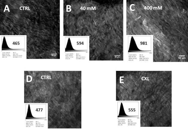
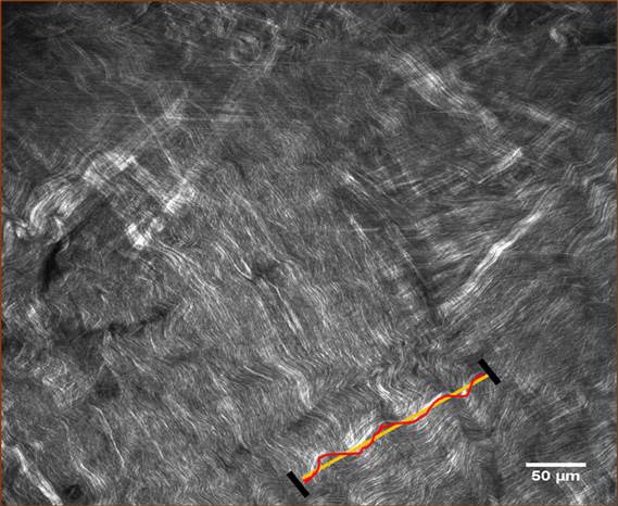
Purpose: Second harmonic generation signals (SHG) are emitted preferentially from collagenous tissue structures and have been used to evaluate photochemically-induced (CXL) crosslinking changes in the cornea. Since therapeutic tissue crosslinking (TXL) using sodium hydroxymethylglycinate (SMG) of the sclera is a potential treatment for high myopia, we explored the use of SHG microscopy to evaluate the effects.

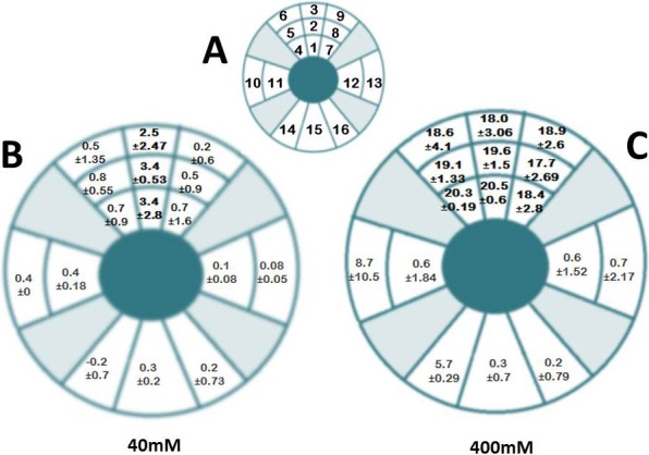
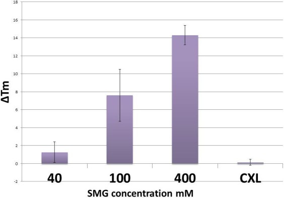
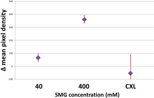
Methods: Single sub-Tenon's (sT) injections (400 μL) using SMG (40-400 mM) were made at the equatorial 12 o'clock position of the right eye of cadaveric rabbit heads (n = 16 pairs). After 3.5 hours, confocal microscopy (CM) was performed using 860 nm two-photon excitation and 400 to 450 nm emission. Pixel density and fiber bundle "waviness" analyses were performed on the images. Crosslinking effects were confirmed using thermal denaturation (Tm) temperature. Comparison experiments with riboflavin photochemical crosslinking were done.

Results: Therapeutic tissue crosslinking localization studies indicated that crosslinking changes occurred at the site of injection and in adjacent sectors. Second harmonic generation signals revealed large fibrous collagenous bundled structures that displayed various degrees of waviness. Histogram analysis showed a nearly 6-fold signal increase in 400 mM SMG over 40 mM. This corresponded to a ΔTm = 13°C for 400 mM versus ΔTm = 4°C for 40 mM. Waviness analysis indicated increased fiber straightening as a result of SMG CXL.

Conclusions: Second harmonic generation signal intensity and fiber bundle waviness is altered by scleral tissue crosslinking using SMG. These changes provide insights into the macromolecular changes that are induced by therapeutic crosslinking technology and may provide a method to evaluate connective tissue protein changes induced by scleral crosslinking therapies.


Figures

Figure 1
Figure 1
Localization of TXL effect sT injection using 40 and 400 mM SMG. A single injection of SMG solution was applied via the sT space using a 25-gauge insulin needle and syringe. Following a 3.5-hour incubation, the tissue was sampled at a series of locations by sectioning the tissue. Each sector was analyzed independently for crosslinking effect by DSC. The corresponding sectors in the contralateral NaHCO3 injected eyes were treated and sectioned in a similar fashion and were expressed as the difference in Tm, or ΔTm. (A) Represents a schematic of the location of injection and excised scleral sectors and is not drawn to scale. The injection site corresponded to sector 2, a 4 × 4 mm area located just anterior to the equator at the 12 o'clock position. Sectors 1 and 3 were approximately 4 × 4 mm square segments taken adjacent to the injection site extending to 5 mm posterior to the limbus anteriorly, and 1.5 cm anterior to the posterior pole posteriorly. These anterior and posterior “margins” represented the extent of tissue sectioning for the remaining sectors as well (4–16). Sectors 4 to 6 were taken from the adjacent superonasal location and sectors 7 to 9 were taken from the adjacent superotemporal location. Sectors 10 and 11 were taken from the medial orbital wall and sectors 12 and 13 were taken from the lateral orbital wall. Sector 15 was taken from the 6 o'clock location with sectors 14 and 16 taken from the adjacent regions. (B) 40 mM SMG and (C) 400 mM SMG are the localization maps for SMG crosslinking. Each sector contains the ΔTm ± SD, representing the shift in denaturation temperature. That is the mean difference in Tm between experimental sets and their corresponding paired controls. The results indicate that a localization of effect occurs in the region of the injection. In 400 mM–treated samples, the localization of effect is similar to 40 mM–treated samples, but encompasses a greater area with significantly greater crosslinking effects, as evidenced by the large shifts in Tm (18.0°–20.5° vs. 2.5°3.4°C). Each value represents the average of a minimum of 3 independent determinations. P values for each sector are not shown but sectors showing statistically significant values were sectors 1 to 3 for 40 mM (B) and 1 to 9 for 400 mM (C).
Figure 2
Figure 2
Concentration-dependent thermal denaturation effects using SMG via sT injection and photochemical crosslinking in ex vivo in situ rabbit eyes. Methods as described previously (see text). The difference in TXL effect taken from the site of injection based on results from Figure 1, varies depending on the concentration of SMG used. No crosslinking effect was observed using the “CXL” riboflavin photochemical crosslinking technique.
Figure 3
Figure 3
Representative images of concentration dependent increases in SHG signal brightness levels produced following induced crosslinking (TXL) using SMG via sT injection and by UVA-riboflavin crosslinking of sclera ex vivo. Each sample has its own paired control. Two different concentration of SMG are shown as (B) 40 mM and (C) 400 mM, as well as (D) riboflavin only control and (E) CXL crosslinking. The absorption spectrum of riboflavin (max ca. 420 nm) is known to overlap with the SHG signal generated (860 nm excitation with 400–450 nm emission) and this could confound the SHG signal although washing the tissue three times in PBS was sufficient to generate baseline signals without artifact (data not shown). The histogram analysis of pixel density accompanies each Figure and is computer software generated (NIS software version 4.3). Statistical comparisons were then performed as shown in Figure 4.
Figure 4
Figure 4
Average of the differences in densitometric SHG signals (mean pixel densities) in scleral intact globes crosslinked via sT injection with 40 and 400 mM SMG solutions, as well as riboflavin photochemical crosslinking (CXL). A nearly 6-fold increase in SHG signal brightness is noted in the 400 mM sample over 40 mM SMG. Photochemically crosslinked samples did not show increased signal as compared to paired control samples.
Figure 5
Figure 5
Representative image and example of fiber waviness analysis (as expressed by linearity). Quantitative collagen fiber waviness analysis was performed with single-focal plane images as described in the text. Treated samples showed 63% and 55% of Waviness-% for 40 mM and 400 mM SMG, respectively (Table).

References

    1. McBrien NA,, Norton TT. Prevention of collagen crosslinking increases form-deprivation myopia in tree shrew. Exp Eye Res. 1994; 59: 475–486. - PubMed
    1. Elsheikh A,, Phillips JR. Is scleral cross-linking a feasible treatment for myopia control? Ophthalmic Physiol Opt 2013; 33: 385–389. - PubMed
    1. Wollensak G. [Friedrich Joseph Haas -- the holy doctor from Moscow]. Klin Monbl Augenheilkd. 2005; 222: 513–515. - PubMed
    1. Wollensak G,, Iomdina E. Long-term biomechanical properties of rabbit sclera after collagen crosslinking using riboflavin and ultraviolet A (UVA). Acta Ophthalmol. 2009; 87: 193–198. - PubMed
    1. Wang M,, Zhang F,, Liu K,, Zhao X. Safety evaluation of rabbit eyes on scleral collagen cross-linking by riboflavin and ultraviolet A. Clin Experiment Ophthalmol. 2015; 43: 156–163. - PubMed

Publication types