. 2017 Jan 5;12(1):e0169584.
doi: 10.1371/journal.pone.0169584. eCollection 2017.

Vascular Endothelial Over-Expression of Human Soluble Epoxide Hydrolase (Tie2-sEH Tr) Attenuates Coronary Reactive Hyperemia in Mice: Role of Oxylipins and ω-Hydroxylases

Affiliations

Vascular Endothelial Over-Expression of Human Soluble Epoxide Hydrolase (Tie2-sEH Tr) Attenuates Coronary Reactive Hyperemia in Mice: Role of Oxylipins and ω-Hydroxylases

Ahmad Hanif et al. PLoS One. .

Abstract

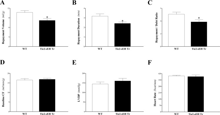
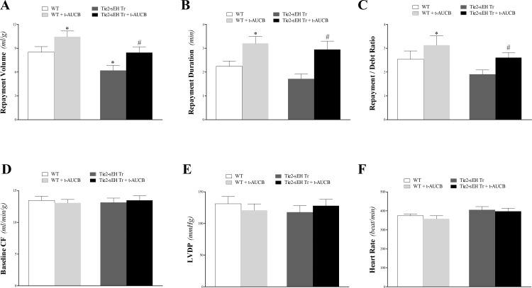
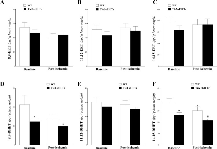
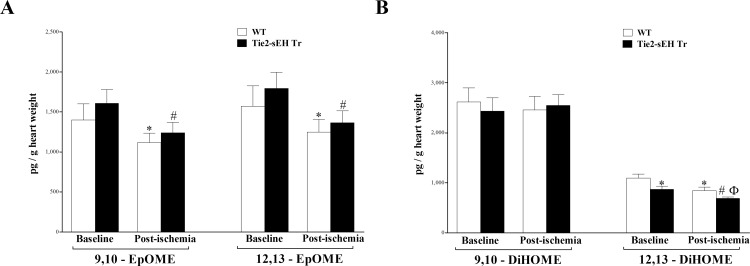
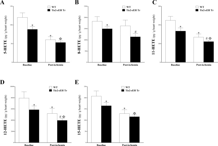
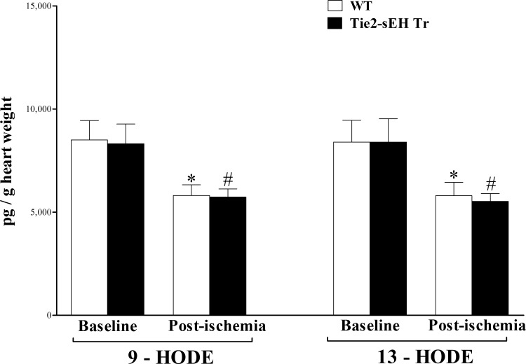
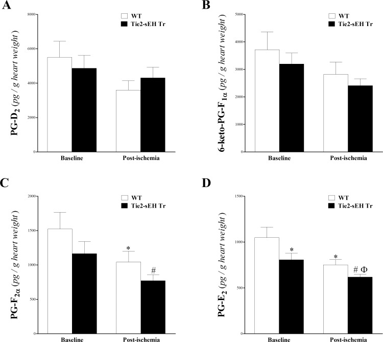
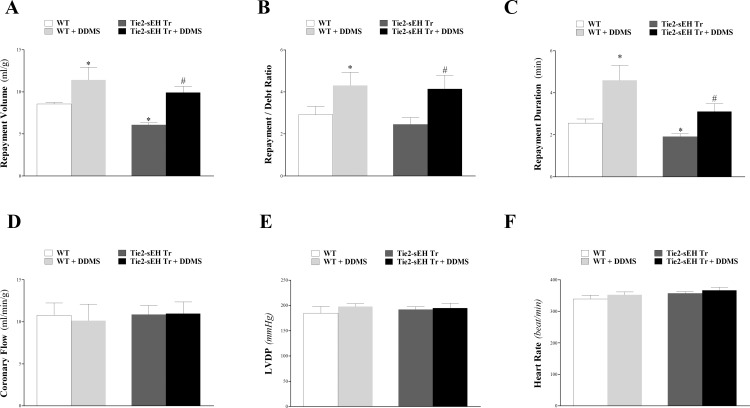
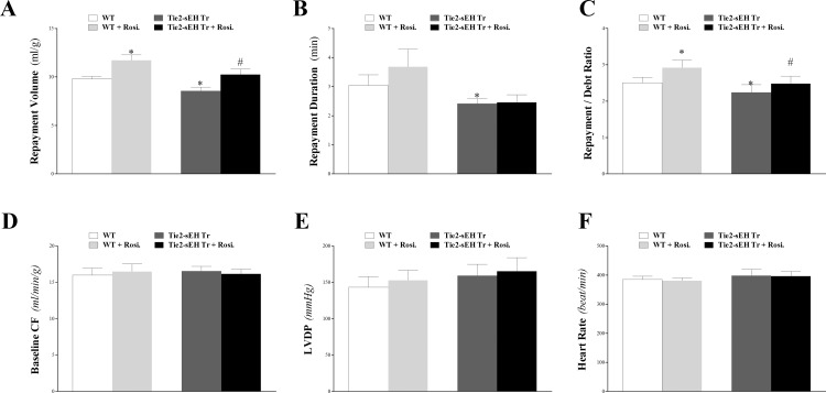
Cytochromes P450 metabolize arachidonic acid (AA) into two vasoactive oxylipins with opposing biologic effects: epoxyeicosatrienoic acids (EETs) and omega-(ω)-terminal hydroxyeicosatetraenoic acids (HETEs). EETs have numerous beneficial physiological effects, including vasodilation and protection against ischemia/reperfusion injury, whereas ω-terminal HETEs induce vasoconstriction and vascular dysfunction. We evaluated the effect of these oxylipins on post-ischemic vasodilation known as coronary reactive hyperemia (CRH). CRH prevents the potential harm associated with transient ischemia. The beneficial effects of EETs are reduced after their hydrolysis to dihydroxyeicosatrienoic acids (DHETs) by soluble epoxide hydrolase (sEH). ω-terminal HETEs are formed by ω-hydroxylase family members. The relationship among endothelial over-expression of sEH (Tie2-sEH Tr), the changes in oxylipins it may produce, the pharmacologic inhibition of ω-hydroxylases, activation of PPARγ, and CRH response to a brief ischemia is not known. We hypothesized that CRH is attenuated in isolated mouse hearts with endothelial sEH over-expression through modulation of oxylipin profiles, whereas both inhibition of ω-hydroxylases and activation of PPARγ enhance CRH. Compared to WT mice, Tie2-sEH Tr mice had decreased CRH, including repayment volume, repayment duration, and repayment/debt ratio (P < 0.05), whereas inhibition of ω-hydroxylases increased these same CRH parameters in Tie2-sEH Tr mice. Inhibition of sEH with t-AUCB reversed the decreased CRH in Tie2-sEH Tr mice. Endothelial over-expression of sEH significantly changed oxylipin profiles, including decreases in DHETs, mid-chain HETEs, and prostaglandins (P < 0.05). Treatment with rosiglitazone, PPARγ-agonist, enhanced CRH (P < 0.05) in both Tie2-sEH Tr and wild type (WT) mice. These data demonstrate that endothelial over-expression of sEH (through changing the oxylipin profiles) attenuates CRH, whereas inhibition of ω-hydroxylases and activation of PPARγ enhance it.


Conflict of interest statement

The authors have declared that no competing interests exist.

Figures

Fig 1
Fig 1. Comparison of coronary reactive hyperemia (CRH) between Tie2-sEH Tr and WT.
Repayment volume (A), repayment duration (B), and repayment/debt ratio (C), were increased in Tie2-sEH Tr compared to WT mice (P < 0.05). Baseline CF (D), LVPD (E), and HR (F) were not different between the two groups. * P < 0.05 versus WT. n = 12 per group.
Fig 2
Fig 2. Effect of the sEH-inhibitor (t-AUCB, 10 μM) on coronary reactive hyperemia (CRH) in Tie2-sEH Tr and WT mice.
The sEH-inhibitor, t-AUCB, enhanced CRH in both Tie2-sEH Tr and WT mice. Repayment volume (A), repayment duration (B), and repayment/debt ratio (C) were increased in both strains. Repayment volume was decreased more in Tie2-sEH Tr compared to WT mice. Baseline CF (D), LVPD (E), and HR (F) were not different between the two groups. * P < 0.05 versus WT. # P < 0.05 versus t-AUCB–treated WT. n = 8 per group.
Fig 3
Fig 3. LC–MS/MS analysis for EETs (8, 9–, 11, 12–, and 14, 15–) and DHETs (8, 9–, 11, 12–, and 14, 15–) levels in WT and Tie2-sEH Tr mouse heart perfusates at baseline and post-ischemia.
8,9-EET (A), 11,12-EET (B), and 14,15-EET (C) were not significantly different between WT and Tie2-sEH Tr mice. Also, the levels of EETs were not affected in response to ischemia in either mouse strain (P > 0.05). 8,9-DHET (D), and 14,15-DHET (F) were decreased in Tie2-sEH Tr compared to WT mice (P < 0.05), whereas 11,12-DHET (E) was not significantly different between the two groups (P > 0.05). Also, 14,15-DHET (F) was decreased in response to ischemia in Tie2-sEH Tr (P < 0.05). * P < 0.05 versus baseline WT. # P < 0.05 versus WT post-ischemia. n = 12 WT and 14 Tie2-sEH Tr.
Fig 4
Fig 4. LC–MS/MS analysis of EpOME and DiHOME levels in WT and Tie2-sEH Tr mouse heart perfusates at baseline and post-ischemia.
9,10- and 12,13-EpOMEs (A) were decreased in response to ischemia in both WT and Tie2-sEH Tr mice (P < 0.05), but were not different between the two groups (P > 0.05). 12, 13-DiHOME (B) level was decreased in Tie2-sEH Tr compared to WT mice (P < 0.05), and was decreased in response to ischemia in both mouse groups (P < 0.05). 9,10-DiHOME level (B) did not change due to ischemia or to sEH endothelial expression (P > 0.05). * P < 0.05 versus baseline WT. # P < 0.05 versus WT post-ischemia. n = 12 WT and 14 Tie2-sEH Tr.
Fig 5
Fig 5. LC–MS/MS analysis of 5-, 8-, 11-, 12- and 15-HETE levels in WT and Tie2-sEH Tr mouse heart perfusates at baseline and post-ischemia.
In Tie2-sEH Tr mice, the levels of 5-HETE (A), 8-HETE (B), 11-HETE (C), 12-HETE (D), 15-HETE (E), were decreased compared to WT mice at baseline and post-ischemia (P < 0.05). In both WT and Tie2-sEH Tr mice, post-ischemic levels of 5-, 11-, 12-, and 15-HETEs were decreased compared to baseline levels (P < 0.05). * P < 0.05 versus baseline WT. # P < 0.05 versus WT post-ischemia. Ф P < 0.05 versus baseline Tie2-sEH Tr. n = 12 WT and 14 Tie2-sEH Tr.
Fig 6
Fig 6. LC–MS/MS analysis of HODEs in WT and Tie2-sEH Tr mouse heart perfusates at baseline and post-ischemia.
In both WT and Tie2-sEH Tr, 9- and 13-HODEs decreased in response to ischemia (P < 0.05). However, they were not significantly different between the two groups at baseline or post-ischemia (P > 0.05). * P < 0.05 versus baseline WT. # P < 0.05 versus WT post-ischemia. n = 12 WT and 14 Tie2-sEH Tr.
Fig 7
Fig 7. LC–MS/MS analysis of 6-keto-PG-F, PG-F, PG-D2, and PG-E2 in WT and Tie2-sEH Tr mouse heart perfusates at baseline and post-ischemia.
The levels of PG-D2 (A) and 6-Keto-PG-F (B) were not significantly changed due to ischemia or between Tie2-sEH Tr and WT mice (P > 0.05). In both Tie2-sEH Tr and WT mice, PG-F (C) was decreased post-ischemia (P < 0.05). PG-E2 (D) was decreased at baseline and post-ischemia in Tie2-sEH Tr compared to WT mice (P < 0.05), and it was also decreased in response to ischemia in Tie2-sEH Tr mice (P < 0.05). * P < 0.05 versus baseline WT. # P < 0.05 versus WT post-ischemia. Ф P < 0.05 versus baseline Tie2-sEH Tr. n = 12 WT and 14 Tie2-sEH Tr.
Fig 8
Fig 8. Effect of the CYP4A-blocker (DDMS, 1 μM) on coronary reactive hyperemia (CRH) in Tie2-sEH Tr and WT mice.
The CYP4A-blocker, DDMS, enhanced CRH in both Tie2-sEH Tr and WT mice. Repayment volume (A), repayment duration (B), and repayment/debt ratio (C) were increased in both strains. Repayment volume was decreased more in Tie2-sEH Tr compared to WT mice. Baseline CF (D), LVPD (E), and HR (F) were not different between the two groups. * P < 0.05 versus WT. # P < 0.05 versus DDMS–treated WT. n = 8 per group.
Fig 9
Fig 9. Effect of the PPARγ-agonist (rosiglitazone, 10 μM) on coronary reactive hyperemia (CRH) in Tie2-sEH Tr and WT mice.
The PPARγ-agonist, rosiglitazone, enhanced CRH in both Tie2-sEH Tr and WT mice. Repayment volume (A), and repayment/debt ratio (C) were increased in both strains. Repayment duration (B) was increased in WT mice, but not in Tie2-sEH Tr mice. Repayment volume was decreased more in Tie2-sEH Tr compared to WT mice. Baseline CF (D), LVPD (E), and HR (F) were not different between the two groups. * P < 0.05 versus WT. # P < 0.05 versus rosiglitazone–treated WT. n = 8 per group.

References

    1. Campbell WB, Gebremedhin D, Pratt PF, Harder DR. Identification of epoxyeicosatrienoic acids as endothelium-derived hyperpolarizing factors. Circ Res. 1996;78(3):415–23. - PubMed
    1. Ellinsworth DC, Shukla N, Fleming I, Jeremy JY. Interactions between thromboxane A(2), thromboxane/prostaglandin (TP) receptors, and endothelium-derived hyperpolarization. Cardiovasc Res. 2014;102(1):9–16. 10.1093/cvr/cvu015 - DOI - PubMed
    1. Catella F, Lawson JA, Fitzgerald DJ, FitzGerald GA. Endogenous biosynthesis of arachidonic acid epoxides in humans: increased formation in pregnancy-induced hypertension. Proc Natl Acad Sci U S A. 1990;87(15):5893–7. - PMC - PubMed
    1. Campbell WB, Deeter C, Gauthier KM, Ingraham RH, Falck JR, Li PL. 14,15-Dihydroxyeicosatrienoic acid relaxes bovine coronary arteries by activation of K(Ca) channels. Am J Physiol Heart Circ Physiol. 2002;282(5):H1656–64. 10.1152/ajpheart.00597.2001 - DOI - PubMed
    1. Larsen BT, Campbell WB, Gutterman DD. Beyond vasodilatation: non-vasomotor roles of epoxyeicosatrienoic acids in the cardiovascular system. Trends Pharmacol Sci. 2007;28(1):32–8. 10.1016/j.tips.2006.11.002 - DOI - PubMed

MeSH terms