. 2017 Jan 5;18(1):15.
doi: 10.1186/s12859-016-1411-0.

Structural prediction of the interaction of the tumor suppressor p27KIP1 with cyclin A/CDK2 identifies a novel catalytically relevant determinant

Affiliations

Structural prediction of the interaction of the tumor suppressor p27KIP1 with cyclin A/CDK2 identifies a novel catalytically relevant determinant

Jinyu Li et al. BMC Bioinformatics. .

Abstract

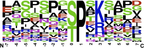
Background: The cyclin-dependent kinase 2 (CDK2) together with its cyclin E and A partners is a central regulator of cell growth and division. Deregulation of CDK2 activity is associated with diseases such as cancer. The analysis of substrates identified S/T-P-X-R/K/H as the CDK2 consensus sequence. The crystal structure of cyclin A/CDK2 with a short model peptide supports this sequence and identifies key interactions. However, CDKs use additional determinants to recognize substrates, including the RXL motif that is read by the cyclin subunits. We were interested to determine whether additional amino acids beyond the minimal consensus sequence of the well-studied substrate and tumor suppressor p27KIP1 were relevant for catalysis.

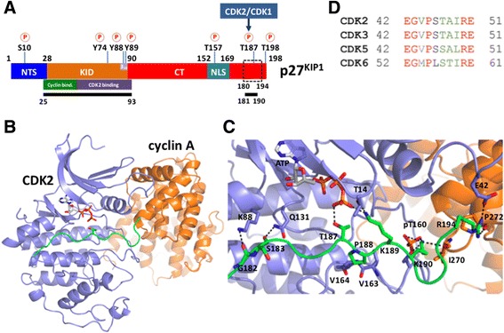
Results: To address whether additional amino acids, close to the minimal consensus sequence, play a role in binding, we investigate the interaction of cyclin A/CDK2 with an in vivo cellular partner and CDK inhibitor p27KIP1. This protein is an intrinsically unfolded protein and, in particular, the C-terminal half of the protein has not been accessible to structural analysis. This part harbors the CDK2 phosphorylation site. We used bioinformatics tools, including MODELLER, iTASSER and HADDOCK, along with partial structural information to build a model of the C-terminal region of p27KIP1 with cyclin A/CDK2. This revealed novel interactions beyond the consensus sequence with a proline and a basic amino acid at the P + 1 and the P + 3 sites, respectively. We suggest that the lysine at P + 2 might regulate the reversible association of the second counter ion in the active site of CDK2. The arginine at P + 7 interacts with both cyclin A and CDK2 and is important for the catalytic turnover rate.

Conclusion: Our modeling identifies additional amino acids in p27KIP1 beyond the consensus sequence that contribute to the efficiency of substrate phosphorylation.

Keywords: Cyclin-dependent kinase; Molecular modeling; Phosphorylation; Tumor suppressor; p27KIP1.


Figures

Fig. 1
Fig. 1
Sequence logo plot represent normalized amino acid frequencies for ±7 amino acids from the phosphorylation site of CDK2 in vivo substrates reported in Table 1. This figure was generated using WebLogo [76]
Fig. 2
Fig. 2
Schematic visualization of p27KIP1 and structural model of the cyclin A/CDK2/p27KIP1 peptide (amino acids 180–194) complex. a Schematic representation of the domain organization of p27KIP1. The N-terminal segment (NTS, residues 1–28), the N-terminal kinase inhibitory domain (KID, residues 28–90), the C-terminal region (CT, residues 91–198), and the nuclear localization sequence (NLS, residues 152–169) are indicated. The known phosphorylation sites observed in p27KIP1 are also indicated [15]. The cyclin and CDK2 binding regions in KID are highlighted [21]. The available crystal structure data for p27KIP1 are highlighted by black bars (PDB ID: 1JSU (residues 25–93) [21] and 2AST (residues 181–190) [29]). The C-terminal binding region (residues 180–194), for which a structural model was constructed, is indicated by a dashed box. b The overall structure is shown in cartoon representation. CDK2, cyclin A, and the peptide substrate (amino acids 180–194 of p27KIP1) are colored in blue, orange, and green, respectively. The model contains ATP. c Close-up view of the binding interface between p27KIP1 and cyclin A/CDK2. Hydrogen bonds are highlighted in black dashed lines. d Sequence alignment showing the conservation of E42 in CDK2 compared with other CDKs. The sequence alignment was obtained using the Clustal Omega webserver [42]. Identical and structurally similar residues are indicated in red and green, respectively
Fig. 3
Fig. 3
Superposition of the predicted model of cyclin A/CDK2/p27KIP1 with cyclin A/CDK2/HHASPRK X-ray structure (PDB ID: 1QMZ [11]). The cartoon representations of CDK2, cyclin A, and the p27KIP1 peptide (amino acids 180–194) are colored in blue, orange, and green, respectively. The peptide substrate in the crystal structure is colored in cyan
Fig. 4
Fig. 4
Superposition of cyclin A/CDK2/p27KIP1 peptide (amino acids 180–194) with the PKA/ATP/2 Mn2+ crystal structure (PDB ID: 1AST [36]). The second Mn2+ in PKA is indicated by cyan sphere. The cartoon representations of CDK2 and p27KIP1 are colored in blue and green, respectively. PKA is colored in cyan. PKA residues are labeled with prime
Fig. 5
Fig. 5
Analysis of cyclin A/CDK2 kinase activity on p27KIP1 substrate peptides. Cyclin A/CDK2 Kinase purified from Sf9 cells was incubated with increasing concentrations of p27KIP1 wild type and the R194A mutant peptide as indicated. Phosphorylation of the peptides was monitored after immobilisation on SAM2 membrane by scintillation counting and analysis by GraphPad Prism software. Shown are mean values ± SD of 5 experiments performed in duplicates
Fig. 6
Fig. 6
Flow chart describing the modeling process as detailed in the Methods section

References

    1. Murray AW. Recycling the cell cycle: cyclins revisited. Cell. 2004;116(2):221–234. doi: 10.1016/S0092-8674(03)01080-8. - DOI - PubMed
    1. Malumbres M, Barbacid M. Cell cycle, CDKs and cancer: a changing paradigm. Nat Rev Cancer. 2009;9(3):153–166. doi: 10.1038/nrc2602. - DOI - PubMed
    1. Zhang J, Yang PL, Gray NS. Targeting cancer with small molecule kinase inhibitors. Nat Rev Cancer. 2009;9(1):28–39. doi: 10.1038/nrc2559. - DOI - PubMed
    1. Malumbres M, Pevarello P, Barbacid M, Bischoff JR. CDK inhibitors in cancer therapy: what is next? Trends Pharmacol Sci. 2008;29(1):16–21. doi: 10.1016/j.tips.2007.10.012. - DOI - PubMed
    1. Chohan TA, Qian H, Pan Y, Chen JZ. Cyclin-dependent kinase-2 as a target for cancer therapy: progress in the development of CDK2 inhibitors as anti-cancer agents. Curr Med Chem. 2015;22(2):237–263. doi: 10.2174/0929867321666141106113633. - DOI - PubMed

LinkOut - more resources