Case Reports
. 2017 Jan 5;17(1):17.
doi: 10.1186/s12885-016-3000-z.

Case report: whole exome sequencing of primary cardiac angiosarcoma highlights potential for targeted therapies

Affiliations
Case Reports

Case report: whole exome sequencing of primary cardiac angiosarcoma highlights potential for targeted therapies

Leah Zhrebker et al. BMC Cancer. .

Abstract

Background: Primary cardiac angiosarcomas are rare, but they are the most aggressive type of primary cardiac neoplasms. When patients do present, it is with advanced pulmonary and/or cardiac symptoms. Therefore, many times the correct diagnosis is not made at the time of initial presentation. These patients have metastatic disease and the vast majority of these patients die within a few months after diagnosis. Currently the treatment choices are limited and there are no targeted therapies available.

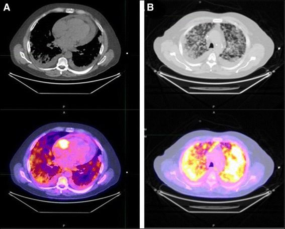
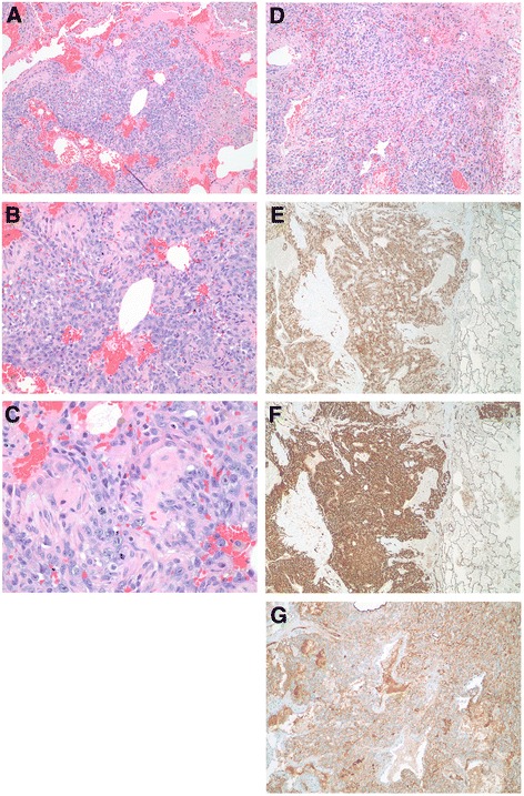
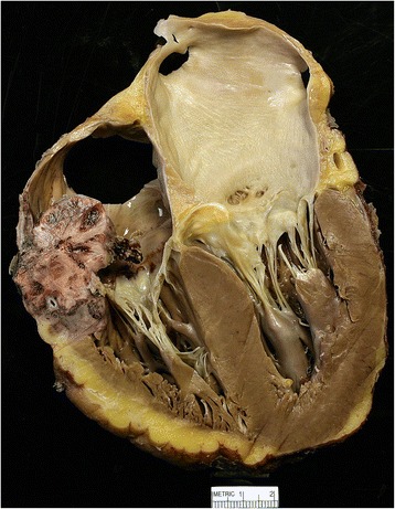
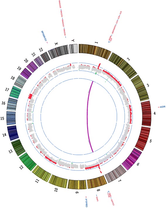
Case presentation: A 56-year-old male presented with shortness of breath, night sweats, and productive cough for a month. Workup revealed pericardial effusion and multiple bilateral pulmonary nodules suspicious for metastatic disease. Transthoracic echocardiogram showed a large pericardial effusion and a large mass in the base of the right atrium. Results of biopsy of bilateral lung nodules established a diagnosis of primary cardiac angiosarcoma. Aggressive pulmonary disease caused rapid deterioration; the patient went on hospice and subsequently died. Whole exome sequencing of the patient's postmortem tumor revealed a novel KDR (G681R) mutation, and focal high-level amplification at chromosome 1q encompassing MDM4, a negative regulator of TP53.

Conclusion: Mutations in KDR have been reported previously in angiosarcomas. Previous studies also demonstrated that KDR mutants with constitutive KDR activation could be inhibited with specific KDR inhibitors in vitro. Thus, patients harboring activating KDR mutations could be candidates for treatment with KDR-specific inhibitors.

Keywords: Activating gene mutation; Cardiac angiosarcoma; Targeted therapies; Whole exome sequencing.


Figures

Fig. 1
Fig. 1
Chest PET/CT Scan. a Chest PET/CT scan of the chest revealed abnormal uptake in the right atrium with an SUV of 14.9. b Chest PET/CT scan showed ground glass appearance and uptake indicative of nodules and pleural effusions in lungs; maximal SUVs of lung nodules were 9.0
Fig. 2
Fig. 2
Images of the tumor from the lung. a-d Hematoxyalin and eosin-stained tumor sections from lung nodules. a Low power magnification (40X) of tumor demonstrated nodules of tumor cells can be seen in a background of abundant, fresh blood cells. b Medium power magnification (100X) showed the tumor cells are both epitheliod and spindle-shaped in appearance. The epitheliod morphology predominates in this area of the tumor. c High power magnification (400X) illustrated the tumor cells have prominent nucleoli. A mitotic figure can be seen in the center of the image confirming the tumor is mitotically active. d Another view of the tumor showing both epitheliod and spindle-shaped tumor cells, but in this section the spindle-shaped cells predominant (40X). e Tumor cells stained positive for CD34, a vascular marker (40X). f Tumor cells showed intense signal for CD31, another vascular marker (40X). g Factor VIII-related antigen, another endothelial marker commonly used to identify vascular tumors, demonstrated positivity in the tumor cells (40X)
Fig. 3
Fig. 3
Cardiac angiosarcoma the right atrium. The tumor was approximately 4 cm in diameter and attached just above the tricuspid valve annulus, extending anteriorly and laterally. The tumor extended into the right atrioventricular sulcus and was present throughout the atrioventricular sulcus shortly after the origin of the right coronary artery and extending posteriorly. For more gross images see Podduturi and Guileyardo [32]
Fig. 4
Fig. 4
Circos plot with whole exome sequencing summary. Copy number alterations (inside track, red = amplifications, green = deletions), Select somatic SNVs (outside track, blue) and structural events (purple lines)

References

    1. Lam KY, Dickens P, Chan AC. Tumors of the heart. A 20-year experience with a review of 12,485 consecutive autopsies. Arch Pathol Lab Med. 1993;117(10):1027–31. - PubMed
    1. Reynen K. Frequency of primary tumors of the heart. Am J Cardiol. 1996;77(1):107. doi: 10.1016/S0002-9149(97)89149-7. - DOI - PubMed
    1. Burke A, Virmani R. More on cardiac myxomas. N Engl J Med. 1996;335(19):1462–3. doi: 10.1056/NEJM199611073351912. - DOI - PubMed
    1. Janigan DT, Husain A, Robinson NA. Cardiac angiosarcomas. A review and a case report. Cancer. 1986;57(4):852–9. doi: 10.1002/1097-0142(19860215)57:4<852::AID-CNCR2820570428>3.0.CO;2-S. - DOI - PubMed
    1. Gal AA, Koss MN, McCarthy WF, Hochholzer L. Prognostic factors in pulmonary fibrohistiocytic lesions. Cancer. 1994;73(7):1817–24. doi: 10.1002/1097-0142(19940401)73:7<1817::AID-CNCR2820730708>3.0.CO;2-K. - DOI - PubMed

Publication types