. 2017 Feb 8:52:61-67.
doi: 10.1016/j.jbiomech.2016.12.008. Epub 2016 Dec 14.

Flow patterns in the jugular veins of pulsatile tinnitus patients

Affiliations

Flow patterns in the jugular veins of pulsatile tinnitus patients

Evan Kao et al. J Biomech. .

Abstract

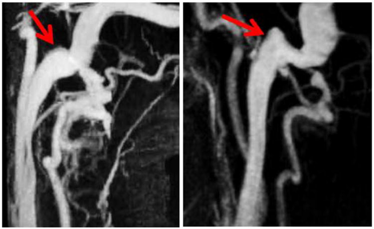
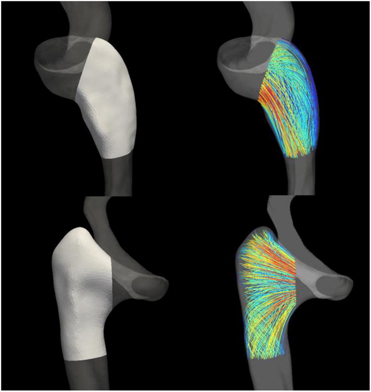
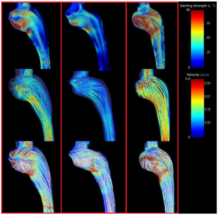
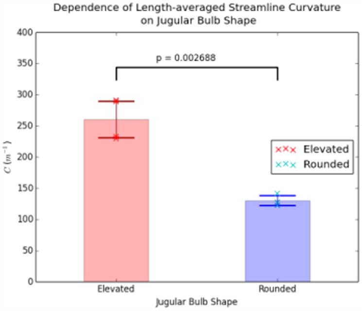
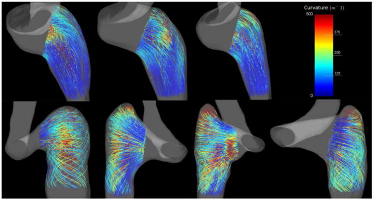
Pulsatile Tinnitus (PT) is a pulse-synchronous sound heard in the absence of an external source. PT is often related to abnormal flow in vascular structures near the cochlea. One vascular territory implicated in PT is the internal jugular vein (IJV). Using computational fluid dynamics (CFD) based on patient-specific Magnetic Resonance Imaging (MRI), we investigated the flow within the IJV of seven subjects, four symptomatic and three asymptomatic of PT. We found that there were two extreme anatomic types classified by the shape and position of the jugular bulbs: elevated and rounded. PT patients had elevated jugular bulbs that led to a distinctive helical flow pattern within the proximal internal jugular vein. Asymptomatic subjects generally had rounded jugular bulbs that neatly redirected flow from the sigmoid sinus directly into the jugular vein. These two flow patterns were quantified by calculating the length-averaged streamline curvature of the flow within the proximal jugular vein: 130.3±8.1m-1 for geometries with rounded bulbs, 260.7±29.4m-1 for those with elevated bulbs (P<0.005). Our results suggest that variations in the jugular bulb geometry lead to distinct flow patterns that are linked to PT, but further investigation is needed to determine if the vortex pattern is causal to sound generation.

Keywords: Computational fluid dynamics; Magnetic resonance imaging; Pulsatile tinnitus.


Conflict of interest statement

Conflict of Interest Statement: There are no conflicts of interest to declare.

Figures

Figure 1
Figure 1
MIP images of the vasculature around the internal jugular vein. The arrows point to the geometry of interest, the jugular bulb, for a rounded bulb (Left) and a raised bulb (Right).
Figure 2
Figure 2
Regions from which streamlines were extracted for calculating the length-averaged streamline curvature (about 1.5 cm along the centerline). These regions were carefully chosen to include only the flow within the proximal internal jugular vein. Top Row: A rounded bulb geometry. Bottom Row: An elevated bulb geometry.
Figure 3
Figure 3
Calculated values of C for specific values of N for a single subject. In this study, N was typically chosen to be in the range of 800-1000 in order to reduce the effect that random streamline seed points had on the value of C.
Figure 4
Figure 4
Simulation results depicting swirling strength and streamlines for all subjects with rounded jugular bulbs. Top Row: Swirling strength distributions throughout the jugular vein representing regions of vortical flow. Note the presence of strong (red) vortex cores in the jugular bulb parallel to flow. Middle Row: Streamlines representing the flow field within the jugular vein. Note that most streamlines from the sigmoid sinus fall straight into the internal jugular vein. Bottom Row: Streamlines visualized with swirling strength. Streamlines are uncolored to emphasize secondary flow structures.
Figure 5
Figure 5
Simulation results depicting swirling strength and streamlines for all subjects with elevated jugular bulbs. Top Row: Swirling strength distributions throughout the jugular vein representing regions of vortical flow. Note the presence of a large vortex core encompassing the nearly the entire volume of the proximal jugular vein. Middle Row: Streamlines representing the flow field within the jugular vein. Note that most streamlines have a significant component that is perpendicular to the direction of flow. Bottom Row: Streamlines visualized with swirling strength. Note the outermost streamlines encircling the prominent vortex core.
Figure 6
Figure 6
Comparison of C for Elevated and Rounded geometries. The cross marks represent values of C for individual subjects, while the transparent bars and associated error bars represent the mean and standard deviation, respectively.
Figure 7
Figure 7
Streamlines in the region of interest colored by curvature. Top Row: Rounded jugular bulbs. Bottom Row: Elevated jugular bulbs.

References

    1. Acevedo-Bolton G, Amans MR, Kefayati S, Halbach V, Saloner D. Four dimensional magnetic resonance velocimetry for complex flow in the jugular vein. Quantitative Imaging in Medicine and Surgery. 2015;5(4):635–7. - PMC - PubMed
    1. Adler JR, Ropper AH. Self-audible venous bruits and high jugular bulbs. Archives of Neurology. 1986;43(3):257–259. - PubMed
    1. Bidhult S, Carlsson M, steding-Ehrenborg K, Arheden H, Heiberg E. A new method for vessel segmentation based on a priori input from medical expertise in cine phase-contrast Magnetic Resonance Imaging. Proceedings of Seventeenth Annual SCMR Scientific Sessions; New Orleans, USA. 2014.
    1. Buckwalter JA, Sasaki CT, Virapongse C, Kier EL, Bauman N. Pulsatile Tinnitus arising from jugular megabulb deformity. Laryngoscope. 1983;93(12):1534–1539. - PubMed
    1. Chanaud RC. Experiments Concerning the Vortex Whistle. The Journal for the Acoustical Society of America. 1963;35(7):953–960.