. 2017 Mar:31:61-70.
doi: 10.1016/j.sleep.2016.07.028. Epub 2016 Nov 10.

Iron and restless legs syndrome: treatment, genetics and pathophysiology

Affiliations

Iron and restless legs syndrome: treatment, genetics and pathophysiology

James R Connor et al. Sleep Med. 2017 Mar.

Abstract

In this article, we review the original findings from MRI and autopsy studies that demonstrated brain iron status is insufficient in individuals with restless legs syndrome (RLS). The concept of deficient brain iron status is supported by proteomic studies from cerebrospinal fluid (CSF) and from the clinical findings where intervention with iron, either dietary or intravenous, can improve RLS symptoms. Therefore, we include a section on peripheral iron status and how peripheral status may influence both the RLS symptoms and treatment strategy. Given the impact of iron in RLS, we have evaluated genetic data to determine if genes are directly involved in iron regulatory pathways. The result was negative. In fact, even the HFE mutation C282Y could not be shown to have a protective effect. Lastly, a consistent finding in conditions of low iron is increased expression of proteins in the hypoxia pathway. Although there is lack of clinical data that RLS patients are hypoxic, there are intriguing observations that environmental hypoxic conditions worsen RLS symptoms; in this chapter we review very compelling data for activation of hypoxic pathways in the brain in RLS patients. In general, the data in RLS point to a pathophysiology that involves decreased acquisition of iron by cells in the brain. Whether the decreased ability is genetically driven, activation of pathways (eg, hypoxia) that are designed to limit cellular uptake is unknown at this time; however, the data strongly support a functional rather than structural defect in RLS, suggesting that an effective treatment is possible.

Keywords: Blood-brain barrier; Dopamine; Genetics; Hypoxia; Iron; RLS.


Figures

Figure 1
Figure 1
CSF ferritin vs serum ferritin for RLS patients (black circles) and controls (open boxes). (adapted from Earley CJ et al NEUROLOGY 2000;54:1698–1700)
Figure 2
Figure 2
Tissue iron concentration at 3 days after tail vein injection of 1000 mg iron isomlatoside or vehicle only compared to animals without any iron deficiency. Note the increased peripheral iron corrects the iron deficiency in the ventral mid-brain and nucleus accumbens without increasing iron in the brain regions without iron deficiency. Iron overload in the brain does not seem to occur. (from Unger, et al, Neuroscience (2013) 246c:179-85)
Figure 3
Figure 3. Striatal microdialysis assessment of extra-cellular non-transferrin bound iron in mice
This figure describes the average iron content from ventral midbrain (VMB) for eight mice with iron isomaltoside 1000 and nine with saline injections. Bars indicate standard errors. Red arrow is the first measure after the injection, which was about 3 h before this measure. Dark periods (18:00–06:00) are indicated by shaded background. Marks at the bottom correspond with Microdialysis samples taken every 3 h starting at 24 h before injection. (from Unger, et al, Neuroscience (2013) 246c:179-85)
Figure 4
Figure 4. MEIS1 is elevated in microvasculature isolated from RLS brains compared to control
These data on 3 samples of RLS and control brain tissue establish that MEIS1 is expressed in brain microvasculature. In combination with the data in Figure 5, the elevation of MEIS1 in RLS is consistent with the concept of brain iron deficiency in RLS. The elevation of MEIS1 is RLS microvasculature is also consistent with activation of hypoxic pathways.
Figure 5
Figure 5. Iron status regulates MEIS1 expression in BREC cultures
These data on 3 sets of BREC cultures demonstrates that iron-deficient BREC cultures have an increased MEIS1 expression while iron-loaded BREC cultures have decreased MEIS1 expression compared to the control cultures.
Figure 6
Figure 6. Alterations in Femoral Artery Blood Flow
This figure demonstrates the femoral artery blood flow in RLS (blue) and control (orange) subjects. Error bars are standard error of the mean. n=18 (9 RLS; 9 age- and gender-matched controls). Baseline femoral artery blood flow is increased 22.5% in RLS subjects. RLS subjects demonstrate only a 1.7% increase in blood flow following exposure to hypoxia, whereas control subjects demonstrate a 7.2% increase in blood flow following hypoxia. (unpublished data)
Figure 7
Figure 7. Alterations in Heart Rate
This figure demonstrates the heart rate measurements in RLS (blue) and control (orange) subjects. As shown in this figure, a trend towards increased baseline heart rate is present in RLS subjects as compared to their age-matched controls. Error bars are standard error of the mean. n=14 (7 RLS; 7 age-matched controls) (unpublished data).
Figure 8
Figure 8. Alterations in Minute Ventilation
This figure demonstrates the minute ventilation measurements in RLS (blue) and control (orange) subjects. Error bars are standard error of the mean. n=14 (7 RLS; 7 age-matched controls). Male RLS subjects had a slightly elevated minute ventilation rate at baseline. Male RLS subjects had a 65% increase in minute ventilation with hypoxic insult.(unpublished data)

References

    1. Hentze MW, et al. Two to tango: regulation of Mammalian iron metabolism. Cell. 2010;142(1):24–38. - PubMed
    1. Muckenthaler MU. How mutant HFE causes hereditary hemochromatosis. Blood. 2014;124(8):1212–1213. - PMC - PubMed
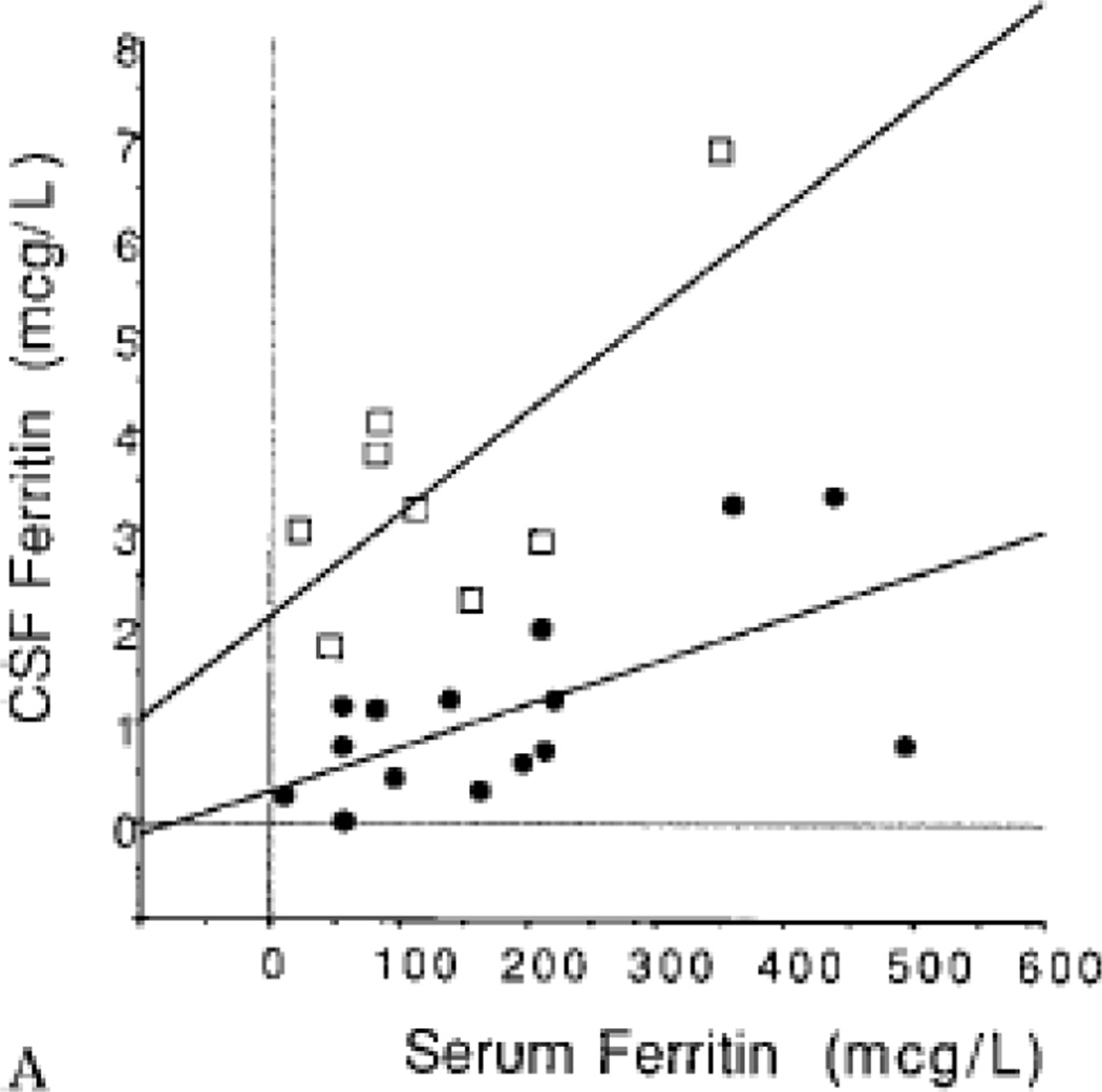
    1. Earley CJ, et al. Abnormalities in CSF concentrations of ferritin and transferrin in restless legs syndrome. Neurology. 2000;54(8):1698–1700. - PubMed
    1. Mizuno S, et al. CSF iron, ferritin and transferrin levels in restless legs syndrome. J Sleep Res. 2005;14(1):43–47. - PubMed
    1. Akyol A, et al. Iron deficiency anemia and restless legs syndrome: is there an electrophysiological abnormality? Clin Neurol Neurosurg. 2003;106(1):23–27. - PubMed

Publication types