Review
. 2017 Feb 10:247:106-126.
doi: 10.1016/j.jconrel.2016.12.032. Epub 2017 Jan 3.

Nanomedicines for dysfunctional macrophage-associated diseases

Affiliations
Review

Nanomedicines for dysfunctional macrophage-associated diseases

Hongliang He et al. J Control Release. .

Abstract

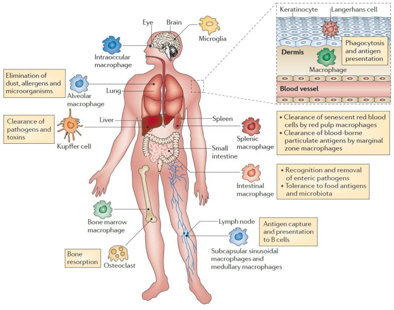
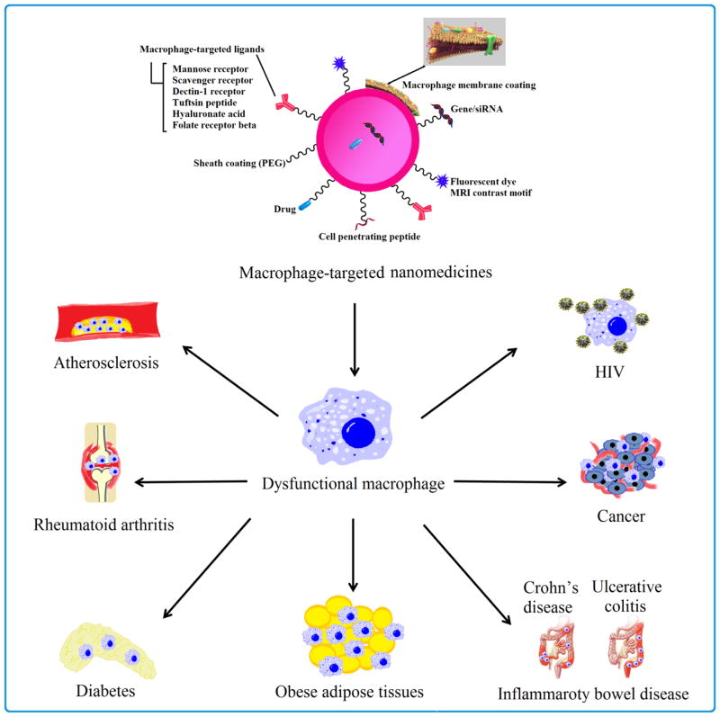
Macrophages play vital functions in host inflammatory reaction, tissue repair, homeostasis and immunity. Dysfunctional macrophages have significant pathophysiological impacts on diseases such as cancer, inflammatory diseases (rheumatoid arthritis and inflammatory bowel disease), metabolic diseases (atherosclerosis, diabetes and obesity) and major infections like human immunodeficiency virus infection. In view of this common etiology in these diseases, targeting the recruitment, activation and regulation of dysfunctional macrophages represents a promising therapeutic strategy. With the advancement of nanotechnology, development of nanomedicines to efficiently target dysfunctional macrophages can strengthen the effectiveness of therapeutics and improve clinical outcomes. This review discusses the specific roles of dysfunctional macrophages in various diseases and summarizes the latest advances in nanomedicine-based therapeutics and theranostics for treating diseases associated with dysfunctional macrophages.

Keywords: Cell receptor; Drug delivery; Gene therapy; In vivo; Macrophage phenotype; Tissue macrophages.


Figures

Fig. 1
Fig. 1
Protective and pathogenic functions of macrophage subsets. Adapted with permission from [4].
Fig. 2
Fig. 2
Graphical illustration of different types of nanoparticles.
Fig. 3
Fig. 3
Multifunctional nanomedicines targeting dysfunctional macrophage-associated diseases.
Fig. 4
Fig. 4
Pro-tumorigenic functions of tumor-associated macrophages. Adapted with permission from [120].
Fig. 5
Fig. 5
Multifunctionality of Man-HA-MnO2 NPs: MnO2 particles (~15 nm) are entrapped in hyaluronic acid with mannan molecules attached. When Man-HA-MnO2 NPs are recognized and uptaken by mannose receptor of TAMs, hyaluronic acid reprograms anti-inflammatory, pro-tumoral M2 TAMs to pro-inflammatory, antitumor M1 macrophages, which might be via a TLR2-MyD88-IRAK1-TRAF6-PKCζ-NK-κB-dependent pathway. The reprogrammed M1 macrophages secrete high level of IL-12, IFN-γ and H2O2. The high reactivity of MnO2 NPs toward H2O2 allows for the simultaneous production of O2 and Mn2+ ions and regulation of pH of tumor hypoxia. Once MnO2 NPs are reduced into Mn2+ ions, they exhibit strong enhancement in both T1- and T2-weighted MRI. Adapted with permission from [130].
Fig. 6
Fig. 6
Scheme of macrophage-membrane-coated emtansine liposome with specific metastasis targeting for suppressing lung metastasis of breast cancer. Macrophage-membrane-coated emtansine liposome was fabricated by coating an emtansine liposome with an isolated macrophage membrane to confer the biomimetic functions of the macrophage, thereby facilitating the specific targeting to metastatic sites and enhancing the therapeutic efficacy on cancer metastasis. Adapted with permission from [89].
Fig. 7
Fig. 7
Potential molecular imaging targets in atherosclerosis. White boxes show putative targets for molecular imaging of atherosclerosis. Atherogenesis involves recruitment of inflammatory cells from blood, represented by the monocyte in the upper-left-hand corner of this diagram. Monocytes are the most numerous leukocytes in atherosclerotic plaque. Recruitment depends on expression of adhesion molecules on macrovascular endothelium, as shown, and on plaque microvessels. Once resident in the arterial intima, activated macrophages become phagocytically active, a process that provides another potential target for plaque imaging. Oxidatively modified low-density lipoprotein–associated epitopes that accumulate in plaques may also serve as targets for molecular imaging. Foam cells may exhibit increased metabolic activity, augmenting their uptake of glucose, a process already measurable in the clinic by 18F-FDG uptake. Activated phagocytes can also elaborate protein-degrading enzymes that can catabolize collagen in the plaque's fibrous cap, weakening it, and rendering it susceptible to rupture and hence thrombosis. Mononuclear phagocytes dying by apoptosis in plaques display augmented levels of phosphatidylserine on their surface. Probes for apoptosis such as annexin V may also visualize complicated atheromata. Microvessels themselves can express not only leukocyte adhesion molecules (shown in green) but also integrins such as αVβ3. Proof-of-principle experiments in animals support each process or molecule in white boxes as target for molecular imaging agents. Adapted with permission from [146].
Fig. 8
Fig. 8
The envisioned paradigm to counteract atherosclerotic plaque development aims to repress lipid-scavenging receptors at the level of lesion-based macrophages. Sugar-based AMs were designed to competitively block oxLDL uptake via binding and regulation of SRs on human macrophages. A library of AMs, with systematic variations in charge, stereochemistry, and sugar backbones, was synthesized and kinetically fabricated into serum-stable, core/shell NPs and screened in vitro to identify shell architectures that exhibited maximal atheroprotective potency. To demonstrate lesion level intervention, the lead NP was administered in an atherosclerosis animal model to challenge lesion development during coronary artery disease. Adapted with permission from [97].
Fig. 9
Fig. 9
Schematic diagram on preparation methods and atherosclerotic lesion targeting property of HA-LT-rHDL. Adapted with permission from [171].
Fig. 10
Fig. 10
Overview of the pathogenesis of rheumatoid arthritis. Monocytes are attracted to the rheumatoid arthritis joint, where they differentiate into macrophages and become activated. They secrete TNF-α and IL-1. TNF-α increases the expression of adhesion molecules on endothelial cells, which recruit more cells to the joint. Chemokines, such as MCP1 and IL-8, are also secreted by macrophages and attract more cells into the joint. IL-1 and TNF induce synovial fibroblasts to express cytokines (such as IL-6), chemokines (such as IL-8), growth factors (such as GM-CSF) and MMPs, which contribute to cartilage and bone destruction. TNF-α contributes to osteoclast activation and differentiation. In addition, IL-1 mediates cartilage degradation directly by inducing the expression of MMPs by chondrocytes. Adapted with permission from [172].
Fig. 11
Fig. 11
Schematic diagram of Notch1 targeting siRNA delivery nanoparticles (siRNA-NPs). (A) The siRNA-NPs are prepared by encapsulating poly-siRNA into tGC nanoparticles. (B) The siRNA-NPs are taken by activated macrophage and the nanoparticles can deliver therapeutic Notch1-specific siRNA in RA treatment. Adapted with permission from [182].
Fig. 12
Fig. 12
Schematic illustration of TPP-PPM/siRNA nanoparticle formation, macrophage-targeting delivery and the release of siRNAs to the cytoplasm. Adapted with permission from [105].
Fig. 13
Fig. 13
Proposed mechanism of dextran–dexamethasone conjugate accumulation in obese VAT, macrophage uptake, and uncoupling of the paracrine loop between M1 macrophages and adipocytes: (a) dextran conjugates (green color) accumulate in the left perirenal adipose tissue (AT) and left gonadal AT after intraperitoneal left-side injection in obese mouse. The anatomical depiction shows mice, which have one mesenteric, two perirenal, and two gonadal AT depots. (b) Transverse cross section of mouse abdomen showing green dextran solution location after administration to the peritoneal cavity. (c) Rapid association of dextran conjugate with M1 macrophages in inflamed AT is enabled by transport across the peritoneum to directly access interstitial cells. (d) Simplified summary of inhibition of paracrine loop between M1 macrophages and adipocytes with dextran–dexamethasone conjugates. In the obese state, hypertrophied adipocytes produce MCP-1, which recruits monocytes, and local release of pro-inflammatory cytokines TNFα and IL-6 induces an M1 macrophage phenotype. M1 macrophages further release pro-inflammatory cytokines and chemokines including MCP-1, TNFα, and IL-6, which further enhance the pro-inflammatory gene expression of adipocytes through NF-κB and further enhance adipocyte inflammation. Dextran conjugates are taken up by M1 macrophages through receptor-mediated endocytosis, and free dexamethasone is released within the cells after esterase hydrolysis. Dexamethasone then binds to the glucocorticoid receptor that inhibits the transcription of pro-inflammatory genes. Additional implicated cells (e.g., T cells) and altered metabolic pathways are not depicted. Adapted with permission from [108].
Fig. 14
Fig. 14
HIV-1-infected macrophages shuttle Nef to B cells to impair follicular IgG2 and IgA responses. Adapted with permission from [198].
Fig. 15
Fig. 15
The concept and rational of targeting HIV-infected cells by CCR5-conjuagted cell derived liposomes. (a) Cell-derived liposomes are prepared by homogenization and extrusion of ghost cells, which membranally express the HIV co-receptor CCR5. The source of the cells can be any autologous lineages that express CCR5 (naturally or exogenously). (b) Cell derived liposomes, which will be targeted against gp120-expressing cells using CCR5 as a targeting ligand, can interfere with all extracellular steps of viral pathogenesis; Such as, syncytium formation between infected cells and uninfected susceptible cells, viral budding from infected cells, viral maturation and attachment to susceptible cells. (c) The cell derived liposomes (when administered along with soluble CD4) are primarily intended to target, fuse with and deliver their cytotoxic content into gp120-expressing/HIV-infected cells. Accordingly, the same mechanism of gp120 binding may also prove efficient in preventing syncytium formation and inactivation of roaming virions by clustering. Adapted with permission from [205].

Similar articles

Cited by

References

    1. Hettinger J, Richards DM, Hansson J, Barra MM, Joschko AC, Krijgsveld J, Feuerer M. Origin of monocytes and macrophages in a committed progenitor. Nature immunology. 2013;14:821–830. - PubMed
    1. Epelman S, Lavine KJ, Randolph GJ. Origin and functions of tissue macrophages. Immunity. 2014;41:21–35. - PMC - PubMed
    1. Wynn TA, Chawla A, Pollard JW. Macrophage biology in development, homeostasis and disease. Nature. 2013;496:445–455. - PMC - PubMed
    1. Murray PJ, Wynn TA. Protective and pathogenic functions of macrophage subsets. Nature reviews immunology. 2011;11:723–737. - PMC - PubMed
    1. Lee KM, Yin C, Verschoor CP, Bowdish DM. Macrophage function disorders. eLS. 2001

Publication types

MeSH terms