. 2017 Mar 2;129(9):1184-1196.
doi: 10.1182/blood-2016-07-728253. Epub 2017 Jan 5.

Murine systemic thrombophilia and hemolytic uremic syndrome from a factor H point mutation

Affiliations

Murine systemic thrombophilia and hemolytic uremic syndrome from a factor H point mutation

Yoshiyasu Ueda et al. Blood. .

Abstract

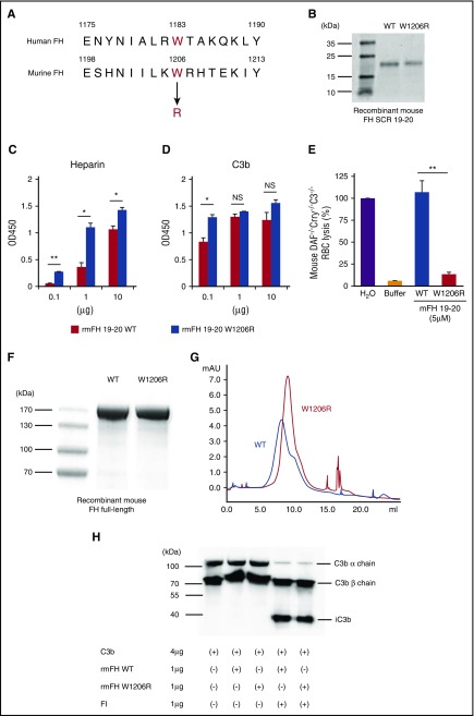
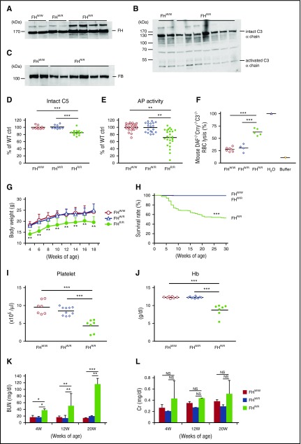
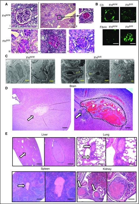
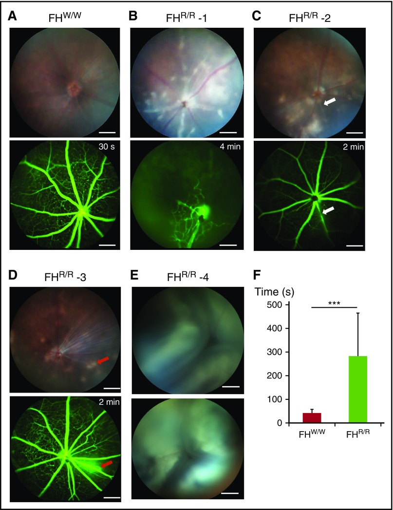
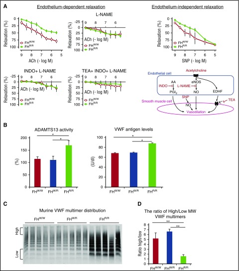
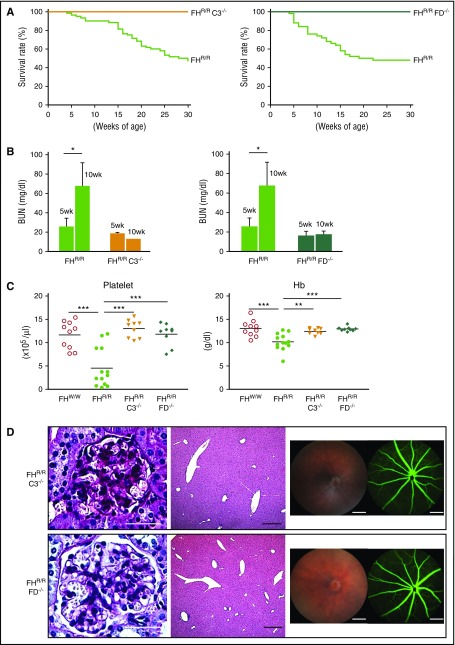
Complement plays a key role in host defense, but its dysregulation can cause autologous tissue injury. Complement activation is normally controlled by regulatory proteins, including factor H (FH) in plasma and membrane cofactor protein (MCP) on the cell surface. Mutations in FH and MCP are linked to atypical hemolytic uremic syndrome, a type of thrombotic microangiopathy (TMA) that causes renal failure. We describe here that disruption of FH function on the cell surface can also lead to disseminated complement-dependent macrovascular thrombosis. By gene targeting, we introduced a point mutation (W1206R) into murine FH that impaired its interaction with host cells but did not affect its plasma complement-regulating activity. Homozygous mutant mice carrying this mutation developed renal TMA as well as systemic thrombophilia involving large blood vessels in multiple organs, including liver, lung, spleen, and kidney. Approximately 30% of mutant mice displayed symptoms of stroke and ischemic retinopathy, and 48% died prematurely. Genetic deficiency of complement C3 and factor D prevented both the systemic thrombophilia and renal TMA phenotypes. These results demonstrate a causal relationship between complement dysregulation and systemic angiopathy and suggest that complement activation may contribute to various human thrombotic disorders involving both the micro- and macrovasculature.


Figures

Figure 1.
Figure 1.
W1206R mutation alters cell surface binding, but not cofactor activity, of mouse FH. (A) Alignment of amino acid sequences showing W1206 of mouse FH is equivalent to W1183 in human FH. (B) Sodium dodecyl sulfate polyacrylamide gel electrophoresis analysis of purified recombinant mouse FH SCR 19-20 and its W1206R variant (Coomassie blue staining). (C) ELISA plate-based heparin and (D) C3b-binding assay of WT and mutant mouse FH SCR 19-20. Heparin and C3b were coated onto plates at 0.1 to 10 μg per well, and recombinant mouse FH SCR 19-20 was added at 0.5 μg per well. (E) When added to 50% mouse serum, WT but not the W1206R mutant form of mouse FH SCR 19-20 caused lysis of complement-susceptible RBCs of DAF−/−Crry−/−C3−/− mice. Hypotonic lysis in pure water was used as reference control (100% lysis). (F) Sodium dodecyl sulfate polyacrylamide gel electrophoresis analysis of purified recombinant full-length mouse FH and its W1206R mutant variant (Coomassie blue). (G) Fast protein liquid chromatography analysis showing that the W1206R mutant FH had strong longer retention on a heparin column than WT mouse FH. Recombinant mouse FH proteins (100 μg) were loaded onto the heparin column in PBS and eluted off with 0.5 M NaCl in PBS. (H) Western blot analysis of products from a cofactor activity assay. The W1206R mutant did not affect the cofactor activity of mouse FH in factor I–mediated cleavage of C3b. Data shown in panels C-E represent mean ± standard deviation (SD) of the results. *P < .05; **P < .01; NS, not significant (Student t test).
Figure 2.
Figure 2.
Complement dysregulation, premature death and development of aHUS symptoms in FH W1206R mutant mice. (A) Western blot analysis of plasma FH levels in WT (FHW/W), heterozygous (FHW/R), and homozygous (FHR/R) mutant mice showing that mutant FH was stable and present at a slightly higher level in FHR/R mice. A polyclonal rabbit anti–mouse FH antibody was used to detect both WT and mutant FH. (B-C) Western blot analysis of plasma C3 (B) and FB (C) levels showing that both intact C3 and FB were lower in FHR/R mice than in FHW/W mice. (D) ELISA of plasma intact C5 showing that FHR/R mice had significantly lower C5 levels than FHW/W or FHW/R mice. (E) ELISA of LPS-induced AP complement activity in 10% mouse sera. Data are normalized to the average value of FHW/W mice (100%). (F) Hemolytic assay using 50% sera from FHW/W, FHW/R, and FHR/R mice and RBCs from DAF−/−Crry−/−C3−/− mice. Hypotonic lysis in pure water was used as reference control (100% lysis). (G) FHR/R mice had lower body weights than FHW/W or FHW/R mice (n = 10 in each group, all male mice). (H) Survival curves of FHW/W (n = 275), FHW/R (n = 310), and FHR/R (n = 147) mice up to 30 weeks of age. (I-J) CBC analysis showing FHR/R mice had thrombocytopenia (I) and anemia with low blood hemoglobin (Hb) levels (J). (K-L) BUN and serum Cr levels in mice at different ages. Serum samples were collected from the same mice at 4, 12, and 20 weeks of age (FHW/W n = 20, FHW/R n = 16, and FHR/R n = 19). Data shown in panels K-L are mean ± SD of the results. Each lane in panels A-C and each symbol in panels D-F,I-J represent an individual mouse (4-25 weeks old). Horizontal bars through the scatterplots in panels D-F,I-J indicate the average values in each group. *P < .05; **P < .01; ***P < .001; NS, not significant (Mantel-Haenszel log-rank test for panel H; 1-way ANOVA and Student t test for other panels).
Figure 3.
Figure 3.
Light, immunofluorescence, and electron microscopy of kidney and other pathology in FH W1206R mutant mice. (A) PAS staining of kidney sections. (i) A normal-looking FHW/W mouse glomerulus. (ii-v) Representative FHR/R mouse kidney sections (from 30 mice analyzed) showed features of aHUS, including arteriolar thrombosis (ii, black square), capillary wall thickening and double contours in glomeruli (iii, white arrows), afferent arteriolar thrombosis in arteriole (iv, yellow asterisk), and expanded matrix and microthrombi in capillary lumen (v, blue arrows). Scale bars represent 25 μm (i-ii, left; iii-v) and 50 μm (ii, right). (B) Immunofluorescence staining of C3 and fibrin or fibrinogen. C3 staining was restricted to the Bowman’s capsule only in FHW/W mice, whereas weak C3 staining of a granular pattern was observed in the glomerulus of FHR/R mice. Strong fibrin and fibrinogen staining of a diffuse pattern was detected both in capillaries and in mesangial lesions of FHR/R mouse glomeruli, but not in FHW/W mouse glomeruli. Scale bar, 25 μm. (C) Electron microscopy of kidneys. A FHW/W mouse (15-week-old female, 1 of 2 WT mice analyzed by electron microscopy) kidney showed normal basement membrane and foot processes. In a representative FHR/R mouse (15-week-old female, 1 of 4 mutant mice analyzed by electron microscopy) kidney, the glomerular capillary wall showed subendothelial expansion with fluffy granular material (yellow arrow) and mesangiolysis with rarefaction of mesangial matrix (red asterisk), with no electron-dense deposits. Scale bars represent 2 μm (FHW/W, left, and FHR/R) and 500 nm (FHW/W, right). (D) hematoxylin and eosin staining of brain sections from a representative FHR/R mouse with neurological abnormalities. Ischemic changes (arrow, left panel), thrombus formation (arrow, right panel) or intracerebral hemorrhage (area circled by dotted line) were observed. Scale bar, 25 μm. (E) Hematoxylin and eosin staining of liver, lung, spleen, and kidney sections from representative FHR/R mice showed thrombi (arrows) in large blood vessels. Scale bars represent 25 μm (left panel of each organ) and 50 μm (right panel of each organ).
Figure 4.
Figure 4.
Retinopathy in FH W1206R mutant mice. (A) A 5-week-old male FHW/W mouse showed normal appearance of retina on funduscopy and rapid dye distribution across retinal blood vessels (<30 seconds) on fluorescein angiography. (B) A 10-week-old male FHR/R mouse exhibited retinal whitening and cotton wool spots by funduscopy and widespread nonperfusion of retinal vasculature, with dilation of the few perfused vessels by fluorescein angiography. This appearance is consistent with a central retinal artery occlusion with limited perfusion provided by a cilioretinal artery. (C) A 5-week-old FHR/R mouse showed retinal whitening by funduscopy and stenosis of vessel (white arrow) by fluorescein angiography. (D) A 10-week-old FHR/R mouse exhibited multiple hypopigmented spots on funduscopy and dye leakage (red arrows) by fluorescein angiography. (E) A 5-week-old male FHR/R mouse had detached retina in both eyes by funduscopy. (F) Time lapse analysis of the arteriovenous phase in fluorescein angiography shows delayed perfusion of retinal blood vessels in FHR/R mice (n = 21) compared with FHW/W mice (n = 10). Data are presented mean ± SD. ***P < .001 (Student t test). In panels B-E, the time label on fluorescein angiography designates the time point at which the picture was taken after dye injection. Scale bars, 200 μm.
Figure 5.
Figure 5.
Vasculopathy and changes in plasma ADAMTS13 and VWF profiles in FH W1206R mutant mice. (A) Endothelium-dependent and independent relaxation of small mesenteric arteries was impaired in FHR/R mice. The endothelium-dependent nature of the phenotype was established by the use of inhibitors of nitric oxide synthase (L-NAME), COX (INDO), and KCa (TEA). An endothelium-independent component of the phenotype was indicated by the use of SNP, which acts directly on vascular smooth muscle cells. Three vessel segments per mouse were analyzed (n = 3 mice). (B) ADAMTS13 activity and plasma VWF levels were significantly higher in FHR/R mice than in FHW/W or FHR/R mice (n = 7 per group for ADAMTS13; n = 3 per group for VWF). (C) Western blot of plasma VWF shows that FHR/R mice had higher levels of total VWF (n = 5; each lane represents 1 mouse) and more abundant lower-molecular-weight (MW) multimers. (D) The ratio of high- to low-molecular-weight VWF multimers, as defined in panel C and quantitated using ImageJ software, was significantly lower in FHR/R mice. Data are from 8-week-old FHW/W, FHW/R, and FHR/R mice. Data in panels B,D represent mean ± SD of results from all mice. *P < .05; **P < .01; ***P < .001 (one-way ANOVA and Student t test). AA, arachidonic acid; eNOS, endothelial nitric oxide synthase; MW, molecular weight; NO, nitric oxide; PGI2, prostaglandin I2.
Figure 6.
Figure 6.
Disease phenotype in FH W1206R mutant mice is mediated by AP complement. (A) FHR/R mice with C3 or FD deficiency survived normally, whereas their FHR/R littermates with normal C3 and FD had high mortality (FHR/RC3−/− n = 64, FHR/RC3+/+ n = 38, FHR/RFD−/− n = 60, and FHR/RFD+/+ n = 36). (B) BUN of FHR/R mice (n = 10) was significantly increased between 5 and 10 weeks of age, whereas BUN in FHR/RC3−/− (n = 6) and FHR/RFD−/− (n = 7) mice remained the same at 5 and 10 weeks of age. (C) CBC analysis showed that FHR/RC3−/− (n = 9) and FHR/RFD−/− (n = 9) mice had normal blood platelet counts and Hb levels, whereas their FHR/R littermates (n = 13) had thrombocytopenia and anemia compared with FHW/W mice (n = 13). (D) Representative pictures of kidney (PAS staining) and liver (hematoxylin and eosin staining) histology, retinal funduscopy, and fluorescence angiography showing that there was no glomerular injury in the kidney or thrombosis in the liver and eye of FHR/RC3−/− and FHR/RFD−/− mice and no retinopathy. Scale bars in panel D represent 25 μm (kidney), 50 μm (liver), and 200 μm (eye). Data in panel B represent mean ± SD. Horizontal bars across scatterplots in panel C represent average values. *P < .05; **P < .01; and ***P < .001 (Mantel-Haenszel log-rank test for panel A; 1-way ANOVA and Student t test for other panels).

Comment in

References

    1. Dunkelberger JR, Song WC. Complement and its role in innate and adaptive immune responses. Cell Res. 2010;20(1):34-50. - PubMed
    1. Walport MJ. Complement. First of two parts. N Engl J Med. 2001;344(14):1058-1066. - PubMed
    1. Rodríguez de Córdoba S, Esparza-Gordillo J, Goicoechea de Jorge E, Lopez-Trascasa M, Sánchez-Corral P. The human complement factor H: functional roles, genetic variations and disease associations. Mol Immunol. 2004;41(4):355-367. - PubMed
    1. Józsi M, Zipfel PF. Factor H family proteins and human diseases. Trends Immunol. 2008;29(8):380-387. - PubMed
    1. Fearon DT. Regulation by membrane sialic acid of beta1H-dependent decay-dissociation of amplification C3 convertase of the alternative complement pathway. Proc Natl Acad Sci USA. 1978;75(4):1971-1975. - PMC - PubMed

Publication types

Substances