. 2017 Jan 1;9(1):124-133.
doi: 10.1093/gbe/evw304.

Extremely Low Genomic Diversity of Rickettsia japonica Distributed in Japan

Affiliations

Extremely Low Genomic Diversity of Rickettsia japonica Distributed in Japan

Arzuba Akter et al. Genome Biol Evol. .

Abstract

Rickettsiae are obligate intracellular bacteria that have small genomes as a result of reductive evolution. Many Rickettsia species of the spotted fever group (SFG) cause tick-borne diseases known as "spotted fevers". The life cycle of SFG rickettsiae is closely associated with that of the tick, which is generally thought to act as a bacterial vector and reservoir that maintains the bacterium through transstadial and transovarial transmission. Each SFG member is thought to have adapted to a specific tick species, thus restricting the bacterial distribution to a relatively limited geographic region. These unique features of SFG rickettsiae allow investigation of how the genomes of such biologically and ecologically specialized bacteria evolve after genome reduction and the types of population structures that are generated. Here, we performed a nationwide, high-resolution phylogenetic analysis of Rickettsia japonica, an etiological agent of Japanese spotted fever that is distributed in Japan and Korea. The comparison of complete or nearly complete sequences obtained from 31 R. japonica strains isolated from various sources in Japan over the past 30 years demonstrated an extremely low level of genomic diversity. In particular, only 34 single nucleotide polymorphisms were identified among the 27 strains of the major lineage containing all clinical isolates and tick isolates from the three tick species. Our data provide novel insights into the biology and genome evolution of R. japonica, including the possibilities of recent clonal expansion and a long generation time in nature due to the long dormant phase associated with tick life cycles.

Keywords: generation time; genome evolution; intra-species genomic diversity; intracellular bacteria; rickettsia.


Figures

<sc>Fig</sc>. 1.—
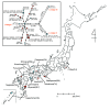
Fig. 1.—
Geographic distribution of Rickettsia japonica analyzed in this study and associated phylogenetic relationships. Isolation locations for the 31 analyzed strains are shown (see Supplementary fig. S2, Supplementary Material online, for more precise information of the strains isolated in Miyazaki prefecture). Clinical and tick isolates are indicated by red and blue circles, respectively. Larger circles that are divided into two or three parts indicate two or three strains isolated from ticks collected in the same location (within a few kilometers) on the same day. The names of the prefecture where the strains were isolated are indicated. The abbreviations of each prefecture are shown in parentheses. In the box, a phylogenetic tree of the 31 R. japonica strains is shown. The tree was constructed by using 112 SNP sites that were identified among the 31 strains. Clinical and tick isolates are indicated by red and blue circles, respectively. Larger circles that are divided into two, three, or four parts represent 2-, 3-, or 4-member strain sets with identical genome sequences in terms of SNPs, respectively. Small dots represent hypothetical intermediates, and the dot-to-dot distance corresponds to one SNP difference. The years and locations of strain isolation are shown in parentheses. Among the three lineages identified, Lineage II contains strains HH-16/17/18, and Lineage III contains one strain (OHH-1). Lineage I contains the other 27 strains. Among the 112 SNPs, we found no homoplasic SNP (the same mutation that occurred in different lineages independently or in parallel).
<sc>Fig</sc>. 2.—
Fig. 2.—
Distribution of SNPs and InDels in the 31 Rickettsia japonica genomes. The top line indicates the full-length R. japonica genome. Genomes from patients and ticks are indicated by red and blue horizontal lines, respectively. Genomic locations of all SNPs and InDels that were identified in each genome are indicated by short vertical lines and triangles, respectively. Synonymous, non-synonymous and intergenic SNPs are indicated by blue, red, and black lines, respectively. Intragenic and intergenic InDels are indicated by equilateral and isosceles triangles, respectively. The dotted rectangles represent the sequence gaps in the rompA gene. SNP clusters found in Lineages II and III are indicated by asterisks. In this figure, HH-13 was used as a reference, and the genomes are ordered according to the maximum likelihood phylogenetic tree shown on the left-hand side.
<sc>Fig</sc>. 3.—
Fig. 3.—
Correlations of SNP distances and geographical or temporal distances among the Lineage I strains. Pair-wise SNP distances and geographical and temporal distances among the Lineage I strains were determined, and the correlations of SNP distances and geographical (left panel) or temporal (right panel) distances were investigated. Lineage I contains two sets of strains (HH-12/13 and HH-16/17/19) that were isolated from ticks that were collected in the same location on the same day. As no SNPs were found in each set of strains, only one strain from each set was included in this analysis. Pearson’s correlation coefficients were calculated using the R package.

Similar articles

Cited by

References

    1. Ando S, Fujita H. 2013. Diversity between spotted fever group rickettsia and ticks as vector (in Japanese; an English abstract available). Med Entomol Zool 64:5–7.
    1. Balashov YS. 1972. Bloodsucking ticks (Ixodoidea)—vectors of diseases of man and animals. Miscellaneous Publications of the Entomological Society of America 8:159–376.
    1. Bennett GM, McCutcheon JP, MacDonald BR, Romanovicz D, Moran NA. 2014. Differential genome evolution between companion symbionts in an insect-bacterial symbiosis. MBio 5:e01697-14. - PMC - PubMed
    1. Blanc G, et al. 2007. Reductive genome evolution from the mother of Rickettsia . PLoS Genet. 3:e14. - PMC - PubMed
    1. Burgdorfer W, Brinton LP. 1975. Mechanisms of transovarial infection of spotted fever Rickettsiae in ticks. Ann N Y Acad Sci. 266:61–72. - PubMed

Publication types