. 2017 Apr;102(4):647-655.
doi: 10.3324/haematol.2016.155812. Epub 2017 Jan 5.

A population of hematopoietic stem cells derives from GATA4-expressing progenitors located in the placenta and lateral mesoderm of mice

Affiliations

A population of hematopoietic stem cells derives from GATA4-expressing progenitors located in the placenta and lateral mesoderm of mice

Ana Cañete et al. Haematologica. 2017 Apr.

Abstract

GATA transcription factors are expressed in the mesoderm and endoderm during development. GATA1-3, but not GATA4, are critically involved in hematopoiesis. An enhancer (G2) of the mouse Gata4 gene directs its expression throughout the lateral mesoderm and the allantois, beginning at embryonic day 7.5, becoming restricted to the septum transversum by embryonic day 10.5, and disappearing by midgestation. We have studied the developmental fate of the G2-Gata4 cell lineage using a G2-Gata4Cre;R26REYFP mouse line. We found a substantial number of YFP+ hematopoietic cells of lymphoid, myeloid and erythroid lineages in embryos. Fetal CD41+/cKit+/CD34+ and Lin-/cKit+/CD31+ YFP+ hematopoietic progenitors were much more abundant in the placenta than in the aorta-gonad-mesonephros area. They were clonogenic in the MethoCult assay and fully reconstituted hematopoiesis in myeloablated mice. YFP+ cells represented about 20% of the hematopoietic system of adult mice. Adult YFP+ hematopoietic stem cells constituted a long-term repopulating, transplantable population. Thus, a lineage of adult hematopoietic stem cells is characterized by the expression of GATA4 in their embryonic progenitors and probably by its extraembryonic (placental) origin, although GATA4 appeared not to be required for hematopoietic stem cell differentiation. Both lineages basically showed similar physiological behavior in normal mice, but clinically relevant properties of this particular hematopoietic stem cell population should be checked in physiopathological conditions.


Figures

Figure 1.
Figure 1.
Analytical flow cytometry analysis of peripheral blood and bone marrow from G2Cre;R26REYFP mice. A: Gating strategy for the identification of different subpopulations of YFP+ cells in lysed peripheral blood B: Gating strategy for the identification of c-Kit+/SCA1+/Lin (KSL) hematopoietic cells in bone marrow (BM). A fraction of the KSL progenitors from bone marrow was YFP+, and 80% of them were Flk2, representing long-term hematopoietic stem cells. Detailed data of these experiments are shown in Table 1. C: The side population of the bone marrow identified by Hoechst 33342 staining includes a fraction of YFP+ cells. D: GATA4 is not expressed in bone marrow (BM) cells by RT-PCR. As a positive control, GATA4 expression was checked in fetal liver (FL) and aorta-gonad-mesonephros tissue (AGM). SSC-A: side scatter; FSC-A: forward scatter; FLK2: fetal liver kinase-2; SP: side population.
Figure 2.
Figure 2.
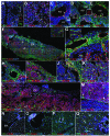
Localization of G2-GATA4 lineage cells (YFP+) in adult and embryonic tissues by confocal microscopy. A,B: YFP+ cells are localized in the bone marrow. Some of these YFP+ cells express c-Kit (arrows in A, separate channels are shown in insert) and CD44 (arrows in B). C–E: YFP+ cells are also localized close to the embryonic aorta (AO) by stage E10. YFP+ cells are abundant around the notochord (NC) and they are also observed in the dorsal mesentery (DM) and in the adjacent coelomic epithelium (CE) (arrowhead in C). Some YFP+ cells can be seen in the aorta, expressing the endothelial marker CD31 (arrow in C). Expression of GATA4 is prominent in the coelomic epithelium, suggesting activity of the enhancer G2 in specific areas of this tissue (D). Some YFP+ cells of the aortic endothelium are GATA4 immunoreactive (arrow and insert in D). The hematopoietic marker RUNX1 is expressed in some YFP+ cells of the aortic endothelium (arrows in E). F–I: YFP+ cells are also abundant in the placenta at stage E12.5. A general view of the placenta (F) shows the distribution of the YFP+ cells, more abundant in the chorionic plate (CP) and around the large vessels (V). The insert shows colocalization with the hematopoietic marker CD41 in the labyrinthine zone (LZ). YFP+ cells are very abundant in the vascular walls, sometimes forming part of the CD31+ endothelium of the large vessels and apparently detaching from it (insert in G). Colocalization with GATA4 and RUNX1 was observed in the vascular walls (arrows in H,I). Note the population of cells expressing GATA4 without activation of the G2 enhancer (arrowhead in H). J,K: YFP+ cells were also found in the fetal liver at stages E11.5 (J) and 13.5 (K). GATA4 is expressed by coelomic epithelium (CE) and mesenchymal cells of the liver, and most of them are YFP+ (arrows in J). Some YFP+ cells also express the hematopoietic marker RUNX1 (arrows in K). L,M: Immunolocalization of GATA2 in the placenta (L) and liver (M) of E11.5 and E12.5 embryos, respectively. No colocalization of GATA2 and YFP is evident in the placenta, but some YFP+ cells are GATA2+ cells in the liver (arrows) and others are not expressing GATA2 (arrowhead). N,O: YFP+ cells in the heart (N) and liver (O) of irradiated mice after four months of injections of bone marrow cells from G2Cre;R26REYFP mice. A number of these cells express the endothelial marker CD31 but they are negative for CD45. Some of these cells are integrated in the endocardium (EN). Clusters of hematopoietic cells appear in the wall of some hepatic sinusoids (arrow in O). P,Q: Busulfan-treated mice injected with cells obtained from G2Cre;R26REYFP embryos. After 4–7 months, putative endothelial CD31+/CD45/YFP+ cells can be seen in the liver after injection of aorta-gonad-mesonephros (P) and placental (Q) cells (arrows). Bars represent 33 μm except for F (200 μm), L (50 μm) and M (25 μm). The images were acquired as 3-channel images by a Leica SP5 II confocal microscope (Leica, Heidelberg, Germany) using LAS AF software and 40X and 63X oil immersion objectives (numerical apertures 1.35 and 1.40, respectively). Levels were adjusted for the entire images using Photoshop 8.0.1.

References

    1. Gao J, Chen YH, Peterson LC. GATA family transcriptional factors: emerging suspects in hematologic disorders. Exp Hematol Oncol. 2015;4:28. - PMC - PubMed
    1. Kuo CT, Morrisey EE, Anandappa R, et al. GATA4 transcription factor is required for ventral morphogenesis and heart tube formation. Genes Dev. 1997;11(8):1048–1060. - PubMed
    1. Molkentin JD, Lin Q, Duncan SA, Olson EN. Requirement of the transcription factor GATA4 for heart tube formation and ventral morphogenesis. Genes Dev. 1997; 11(8):1061–1072. - PubMed
    1. Narita N, Bielinska M, Wilson DB. Wild-type endoderm abrogates the ventral developmental defects associated with GATA-4 deficiency in the mouse. Dev Biol. 1997;189(2):270–274. - PubMed
    1. Rojas A, De Val S, Heidt AB, Xu SM, Bristow J, Black BL. Gata4 expression in lateral mesoderm is downstream of BMP4 and is activated directly by Forkhead and GATA transcription factors through a distal enhancer element. Development. 2005;132(15):3405–3417. - PubMed

Publication types

MeSH terms