. 2017 Mar 1;28(5):587-599.
doi: 10.1091/mbc.E16-07-0555. Epub 2017 Jan 5.

Insulin signaling regulates a functional interaction between adenomatous polyposis coli and cytoplasmic dynein

Affiliations

Insulin signaling regulates a functional interaction between adenomatous polyposis coli and cytoplasmic dynein

Feng J Gao et al. Mol Biol Cell. .

Abstract

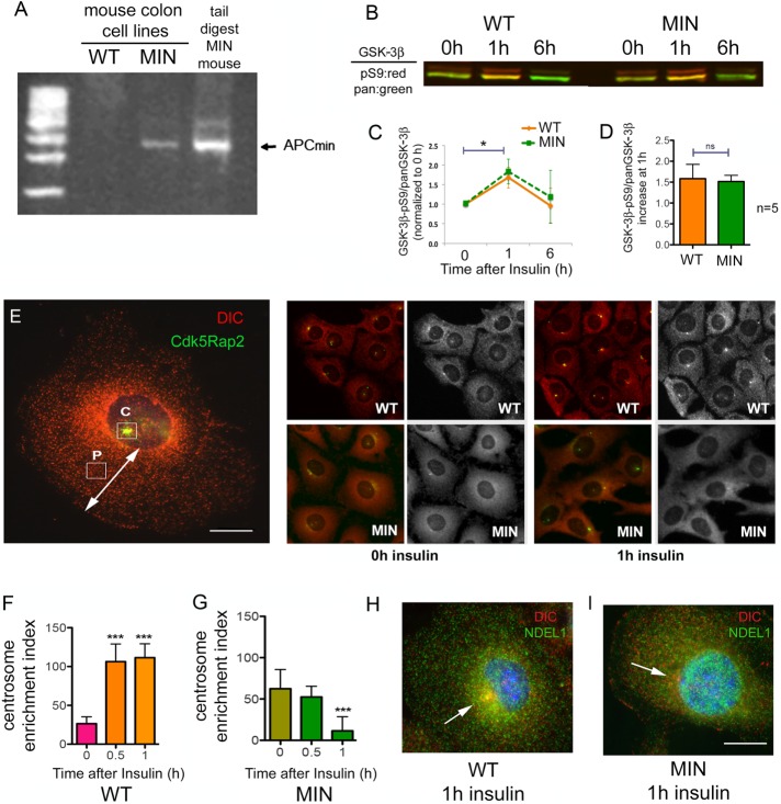
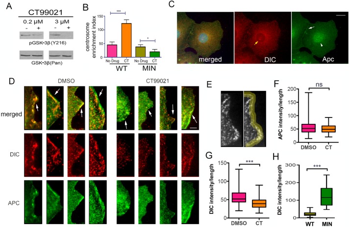
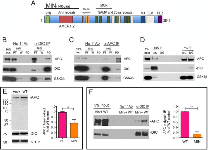
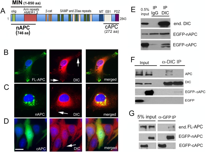
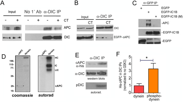
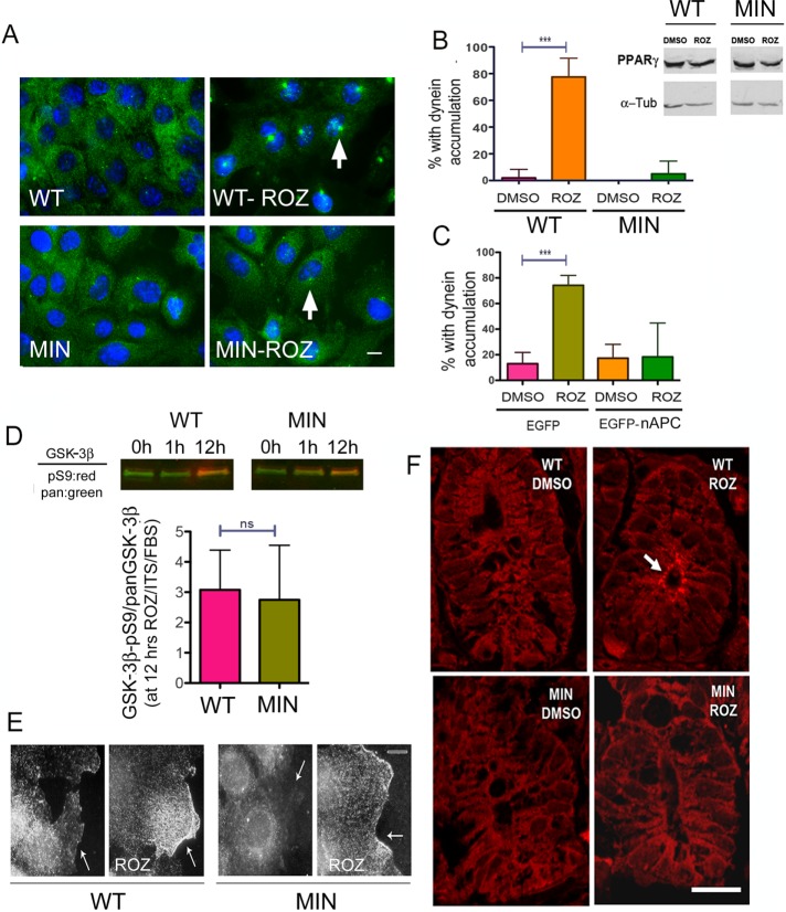
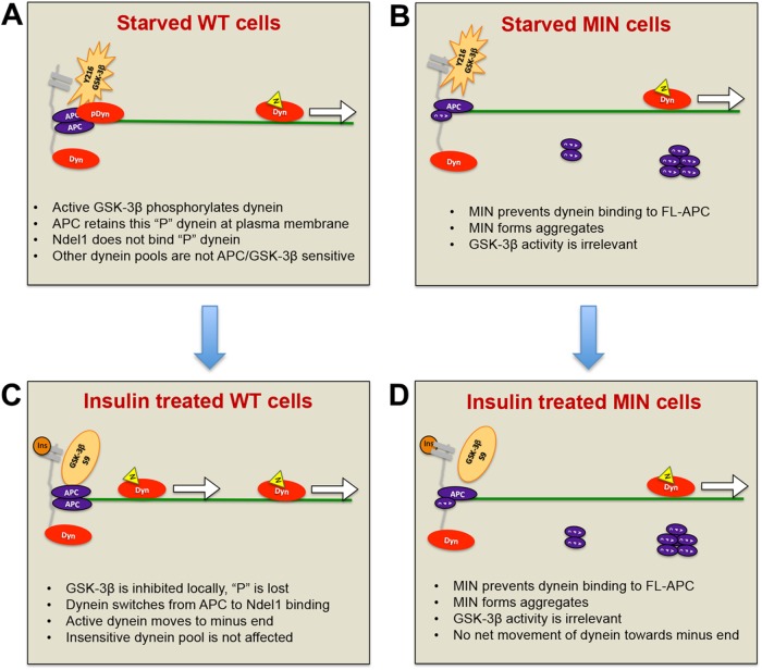
Diabetes is linked to an increased risk for colorectal cancer, but the mechanistic underpinnings of this clinically important effect are unclear. Here we describe an interaction between the microtubule motor cytoplasmic dynein, the adenomatous polyposis coli tumor suppressor protein (APC), and glycogen synthase kinase-3β (GSK-3β), which could shed light on this issue. GSK-3β is perhaps best known for glycogen regulation, being inhibited downstream in an insulin-signaling pathway. However, the kinase is also important in many other processes. Mutations in APC that disrupt the regulation of β-catenin by GSK-3β cause colorectal cancer in humans. Of interest, both APC and GSK-3β interact with microtubules and cellular membranes. We recently demonstrated that dynein is a GSK-3β substrate and that inhibition of GSK-3β promotes dynein-dependent transport. We now report that dynein stimulation in intestinal cells in response to acute insulin exposure (or GSK-3β inhibition) is blocked by tumor-promoting isoforms of APC that reduce an interaction between wild-type APC and dynein. We propose that under normal conditions, insulin decreases dynein binding to APC to stimulate minus end-directed transport, which could modulate endocytic and secretory systems in intestinal cells. Mutations in APC likely impair the ability to respond appropriately to insulin signaling. This is exciting because it has the potential to be a contributing factor in the development of colorectal cancer in patients with diabetes.


Figures

FIGURE 1:
FIGURE 1:
MIN cells respond differently to acute insulin exposure or GSK-3β inhibition after 12 h in starvation medium. (A) Primers for genotyping APC+/MIN mice were used to determine that the cell line derived from APC+/min mouse colon (MIN), but not the cell line derived from WT mice, expresses the truncated MIN isoform of APC. Sequencing indicated that the MIN cells retain one copy of the WT APC gene (unpublished data). (B) The ratio of inactivated GSK-3β (GSK-3β-pS9) to total GSK-3β (pan–GSK-3β) for WT and MIN cells exposed to 0, 1, or 6 h of insulin was determined by Western blot. Phospho-S9 antibody band, red; pan–GSK-3β band, green. (C) Li-Cor densitometry analysis of three Western blots shows mean ± 95% CI. *p < 0.05 by one-way ANOVA. (D) Average increase in S9/pan–GSK-3β after 1 h was determined from five Western blots. (E) Cdk5RAP2 IF (green) was used to label centrosomes in WT and MIN cells costained for DIC (red). Left, CEI calculated by subtracting the intensity of DIC fluorescence measured at a site halfway between the nucleus and the cell periphery (P) from the DIC intensity measured at the centrosome (C). Scale bar, 10 µm. Right, representative WT and MIN cells before and after insulin. The grayscale shows DIC only. (F, G) Acute insulin exposure to starved cells increased CEI in WT cells but not in MIN cells. Significance in C and D was determined with two-tailed paired Student’s t test from three (C) or five (D) separate experiments. Significance in F and G was determined by ANOVA from four independent experiments, ∼500 cells/condition. ***p < 0.001. (H) A WT cell (left) exposed to insulin for 1 h shows accumulation of both DIC (red) and Ndel1 (green) at the centrosome. (I) This was not the case in MIN cells. Scale bar, 10 µm.
FIGURE 2:
FIGURE 2:
Microtubule organization is similar in WT and MIN cells with and without 1-h insulin exposure. Normal full culture medium was replaced with serum- and insulin-free medium for 12 h, and then insulin (ITS, 10 µM) was added for 1 h to one set of cultures. (A) WT cells and (B) MIN cells with no added insulin or (C) WT cells and (D) MIN cells that were exposed to insulin for 1 h were fixed and processed for α-tubulin IF. Insets, individual cells at higher magnification (63×). Scale bars, 50 µm (20× image), 10 µm (inset).
FIGURE 3:
FIGURE 3:
Dynein is reduced at the cell periphery in WT cells. (A) A GSK-3β inhibitor, CT99021 (CT), prevents an activating tyrosine (auto) phosphorylation (Y216). (B) Acute GSK-3β inhibition with CT also increased CEI in WT but not MIN cells. Significance determined by ANOVA from four independent experiments, ∼500 cells/condition. *p < 0.05, ***p < 0.001 (C) Punctate APC (green) and DIC (red) IF occur throughout the cytoplasm in a WT cell. DIC can be seen at the centrosome (yellow arrow, middle). APC is more pronounced at cell junctions (white arrow, right) and in the nucleus (white arrowhead, right). Scale bar, 10 µm. (D) Digital enlargements of four WT cells treated with CT99021 for 1 h (right) and 4 untreated cells (left) reveal some overlap of DIC and APC at leading edges of the cell periphery (arrows). Scale bar, 5 µm. (E) Pixel intensities were measured along a 5-µm-wide line drawn at the cell peripheries of 74 interphase cells for each condition. (F, G) CT99021 reduced the average DIC pixel intensity but not the average APC intensity at the cell periphery. (H) Acute insulin exposure reduced the peripheral DIC intensity in WT but not MIN cells. Significance in F–H was determined using an unpaired two-tailed Student’s t test from three independent experiments; 75 cells/condition. ***p < 0.001.
FIGURE 4:
FIGURE 4:
The MIN isoform interferes with an interaction between dynein and APC. (A) Domains in full-length APC and MIN APC. olig, oligomerization domain; Arm (armadillo) repeats interact with AMER membrane-associated proteins; 15–amino acid repeats are β-catenin–binding domains; MCR, mutation cluster region; SAMP and 20–amino acid repeats are important for Wnt signaling; MT, a basic region that confers microtubule binding; EB1, EB1-binding domain; PDZ, binding domain for PDZ-containing proteins. (B) Dynein was immunoprecipitated from WT mouse brain extract using the DIC 74.1 antibody. Both APC and GSK-3β were present in the immunoprecipitate but not in the no-antibody control (No 1˚ antibody). FT, flowthrough, that is, material that did not bind in the IP. W, material that was released from the IP in the first wash. (C) DIC and GSK-3β also coimmunoprecipitated specifically with APC pulled down by the APC-M2 antibody. (D) The dynein IP was repeated using mouse IgG as a nonspecific antibody control. Endogenous APC and GSK-3β coprecipitated specifically with the DIC immunoprecipitate but not the IgG immunoprecipitate. (E) By Western blotting, MIN brain extracts contain ∼50% of the full-length APC in WT extracts, as quantified from four separate experiments. Significance was determined by paired two-tailed Student’s t test, **p < 0.01. The smaller MIN isoform is not detected with the APC-M2 antibody. DIC and α-tubulin (α-Tub) levels do not appear to be different. (F) Significantly less full-length APC coimmunoprecipitates with dynein from MIN brain extract. The amount of APC in DIC immunoprecipitates from WT and MIN brain extract was quantified from three separate experiments. Significance was determined by paired two-tailed Student’s t test, **p < 0.01.
FIGURE 5:
FIGURE 5:
A C-terminal APC fragment expressed in Cos-7 cells coimmunoprecipitates with endogenous dynein. (A) Location of an N-terminal 746–amino acid APC fragment (nAPC) and a C-terminal 272–amino acid APC (cAPC) fragment used in several experiments. Also shown is the 850–amino acid MIN isoform. (B) Full-length APC fused to EGFP (green) transiently expressed in Cos-7 cells localizes along MTs in cellular protrusions. DIC (red) is concentrated in the cytoplasm but present near the peripheral regions of protrusions (arrows). (C) EGFP-nAPC (green) is enriched in large aggregates in the cytoplasm. Very small puncta of EGFP-nAPC associate with DIC (red) in cellular protrusions at the cell periphery (arrow). (D) EGFP-cAPC (green) is diffuse throughout the cytoplasm and is present in the nucleus. Scale bar, 10 µm. (E) Western blot of proteins in a dynein immunoprecipitate. Top, endogenous dynein (end. DIC) is precipitated by a dynein antibody but not by nonspecific mouse IgG. Middle, EGFP-nAPC is present in both the IgG and the DIC immunoprecipitate. Bottom, EGFP-cAPC coprecipitated specifically with dynein, not IgG. The lower anti-GFP–labeled band may be a proteolytic fragment or a modified peptide that is enriched in the DIC immunoprecipitate. (F) Transient expression of EGFP-cAPC, but not EGFP alone, reduced the amount of endogenous full-length APC (APC) that coprecipitated with dynein. (G) Endogenous APC (end. FL-APC) did not coprecipitate with EGFP-cAPC but did coprecipitate with EGFP-nAPC, which contains an oligomerization domain.
FIGURE 6:
FIGURE 6:
The interaction of dynein with the C-terminus of APC is modulated by GSK-3β activity and dynein phosphorylation. (A) A GSK-3β inhibitor CT99021 (CT) reduced the amount of endogenous APC that coprecipitated with endogenous dynein. (B) Similarly, less EGFP-cAPC coprecipitated with dynein from cells exposed to CT. (C) An EGFP antibody was used to pull down transiently expressed, EGFP-tagged dynein subunit IC1B. More endogenous APC coprecipitated with a WT construct than with an IC1B construct engineered with nonphosphorylatable S87A and T88V mutations in key GSK-3β sites, EGFP-IC1B (M). (D) Left, Coomassie-stained gel of histidine-tagged cAPC (cAPC) or purified bovine brain dynein (dynein). Both were exposed to purified GSK-3β in an in vitro kinase assay. Right, autoradiogram (autorad) shows γ-32P-ATP incorporation in several dynein subunits but not in cAPC. HC, heavy chain; IC, intermediate chain; LIC, light intermediate chain. (E) His-tagged cAPC preferentially coprecipitated with dynein that had been previously phosphorylated by GSK-3β. (F) Quantitation of three separate IPs, average ± 95% CI. Significance was determined by paired two-tailed Student’s t test, *p < 0.05.
FIGURE 7:
FIGURE 7:
The MIN mutation alters the dynein distribution response to rosiglitazone (ROZ) in vivo. (A, B) A 12-h exposure of starved cells to serum plus ITS and ROZ causes DIC (green) to accumulate at centrosomes in WT but not MIN cells. Treatment of WT or MIN cells with ROZ does not lead to an increase in PPARγ expression (top, Western blots). (C) ROZ-induced dynein accumulation is also defective if EGFP-nAPC isoform is transiently expressed in HCT116 cells. (D) Inhibition of GSK-3β is detected after 12 h of ROZ treatment in both WT and MIN cells. (E) Phospho-AKT immunoreactivity is observed at the cell periphery (arrows) in both cell types after ROZ treatment. (F) Colonic crypts from WT but not MIN mice showed an alteration in DIC distribution (red) after 6 d of gavage-administered rosiglitazone. Significant differences in B and C were determined by ANOVA from three separate experiments, 75 cells/treatment; ***p < 0.001. Lack of a significant difference in D was determined by the paired two-tailed Student’s t test from five experiments. ns, not significantly different.
FIGURE 8:
FIGURE 8:
Model for how APC mutations affect insulin-induced dynein movement toward MT minus ends. (A) WT cells in the absence of insulin or serum factors: GSK-3β (orange star) near the membrane (gray) is phosphorylated on Y216 and active. Nearby dynein motors are phosphorylated, rendering them more likely to bind to APC than to Ndel1. Here phosphorylated dynein (pDyn, red oval) is shown bound to an APC homodimer (purple ovals) at sites of MT (green line) capture at the plasma membrane. Unphosphorylated dynein (Dyn) may be bound to Ndel1 (yellow triangle) and actively translocating (arrow). (B) Insulin (dark orange circle) binds to receptors, activating a signaling pathway that locally and transiently inactivates GSK-3β by S9 phosphorylation. Dynein becomes locally and transiently dephosphorylated, is released from APC, binds to Ndel1, and then moves toward MT minus ends. The increase in number of processive dyneins results in minus-end accumulation at centrosomes (not shown). (C) MIN cells in the absence of insulin or serum factors: GSK-3β is active, but there is a reduced pool of dynein associated with FL-APC because MIN APC blocks the interaction. MIN APC also forms aggregates in the cytoplasm. (D) Normal inhibition of GSK3β occurs in response to insulin signaling but as the APC-bound, insulin-sensitive pool of dynein is reduced, the net effect of insulin on motor distribution is minimal.

Similar articles

Cited by

References

    1. Adames NR, Cooper JA. Microtubule interactions with the cell cortex causing nuclear movements in Saccharomyces cerevisiae. J Cell Biol. 2000;149:863–874. - PMC - PubMed
    1. Ait-Omar A, Monteiro-Sepulveda M, Poitou C, Le Gall M, Cotillard A, Gilet J, Garbin K, Houllier A, Chateau D, Lacombe A, et al. GLUT2 accumulation in enterocyte apical and intracellular membranes: a study in morbidly obese human subjects and ob/ob and high fat-fed mice. Diabetes. 2011;60:2598–2607. - PMC - PubMed
    1. Allan VJ. Cytoplasmic dynein. Biochem Soc Trans. 2011;39:1169–1178. - PubMed
    1. Anderson CB, Neufeld KL, White RL. Subcellular distribution of Wnt pathway proteins in normal and neoplastic colon. Proc Natl Acad Sci USA. 2002;99:8683–8688. - PMC - PubMed
    1. Andres SF, Santoro MA, Mah AT, Keku JA, Bortvedt AE, Blue RE, Lund PK. Deletion of intestinal epithelial insulin receptor attenuates high-fat diet-induced elevations in cholesterol and stem, enteroendocrine, and Paneth cell mRNAs. Am J Physiol Gastrointest Liver Physiol. 2015;308:G100–G111. - PMC - PubMed

MeSH terms