

From Microscale Devices to 3D Printing: Advances in Fabrication of 3D Cardiovascular Tissues
- PMID: 28057791
- PMCID: PMC5224928
- DOI: 10.1161/CIRCRESAHA.116.308538
From Microscale Devices to 3D Printing: Advances in Fabrication of 3D Cardiovascular Tissues
Abstract
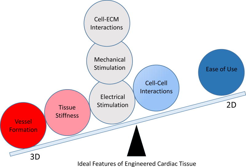
Current strategies for engineering cardiovascular cells and tissues have yielded a variety of sophisticated tools for studying disease mechanisms, for development of drug therapies, and for fabrication of tissue equivalents that may have application in future clinical use. These efforts are motivated by the need to extend traditional 2-dimensional (2D) cell culture systems into 3D to more accurately replicate in vivo cell and tissue function of cardiovascular structures. Developments in microscale devices and bioprinted 3D tissues are beginning to supplant traditional 2D cell cultures and preclinical animal studies that have historically been the standard for drug and tissue development. These new approaches lend themselves to patient-specific diagnostics, therapeutics, and tissue regeneration. The emergence of these technologies also carries technical challenges to be met before traditional cell culture and animal testing become obsolete. Successful development and validation of 3D human tissue constructs will provide powerful new paradigms for more cost effective and timely translation of cardiovascular tissue equivalents.
Keywords: biocompatible materials; heart; printing, three-dimensional; stem cells; tissue engineering.
© 2017 American Heart Association, Inc.
Conflict of interest statement
DISCLOSURES None.
Figures
Similar articles
-
Advances in three-dimensional bioprinted stem cell-based tissue engineering for cardiovascular regeneration.J Mol Cell Cardiol. 2022 Aug;169:13-27. doi: 10.1016/j.yjmcc.2022.04.017. Epub 2022 May 12. J Mol Cell Cardiol. 2022. PMID: 35569213 Free PMC article. Review.
-
Advances in tissue engineering of vasculature through three-dimensional bioprinting.Dev Dyn. 2021 Dec;250(12):1717-1738. doi: 10.1002/dvdy.385. Epub 2021 Jul 2. Dev Dyn. 2021. PMID: 34115420 Review.
-
3D Printing of Tissue Engineered Constructs for In Vitro Modeling of Disease Progression and Drug Screening.Ann Biomed Eng. 2017 Jan;45(1):164-179. doi: 10.1007/s10439-016-1640-4. Epub 2016 May 11. Ann Biomed Eng. 2017. PMID: 27169894 Free PMC article. Review.
-
Advances in Translational 3D Printing for Cartilage, Bone, and Osteochondral Tissue Engineering.Small. 2022 Sep;18(36):e2201869. doi: 10.1002/smll.202201869. Epub 2022 Jun 17. Small. 2022. PMID: 35713246 Review.
-
Unveiling the potential of melt electrowriting in regenerative dental medicine.Acta Biomater. 2023 Jan 15;156:88-109. doi: 10.1016/j.actbio.2022.01.010. Epub 2022 Jan 10. Acta Biomater. 2023. PMID: 35026478 Free PMC article. Review.
Cited by
-
Reconstructing the heart using iPSCs: Engineering strategies and applications.J Mol Cell Cardiol. 2021 Aug;157:56-65. doi: 10.1016/j.yjmcc.2021.04.006. Epub 2021 Apr 22. J Mol Cell Cardiol. 2021. PMID: 33895197 Free PMC article. Review.
-
3D Bioprinting of cardiac tissue and cardiac stem cell therapy.Transl Res. 2019 Sep;211:64-83. doi: 10.1016/j.trsl.2019.04.004. Epub 2019 Apr 20. Transl Res. 2019. PMID: 31078513 Free PMC article. Review.
-
3D vector field-guided toolpathing for 3D bioprinting.Commun Eng. 2025 Aug 14;4(1):154. doi: 10.1038/s44172-025-00489-0. Commun Eng. 2025. PMID: 40813482 Free PMC article.
-
Advances in 3D Bioprinting: Techniques, Applications, and Future Directions for Cardiac Tissue Engineering.Bioengineering (Basel). 2023 Jul 16;10(7):842. doi: 10.3390/bioengineering10070842. Bioengineering (Basel). 2023. PMID: 37508869 Free PMC article. Review.
-
Stem Cells and Extrusion 3D Printing for Hyaline Cartilage Engineering.Cells. 2020 Dec 22;10(1):2. doi: 10.3390/cells10010002. Cells. 2020. PMID: 33374921 Free PMC article. Review.
References
-
- Plunkett N, O’Brien FJ. Bioreactors in tissue engineering. Technol Health Care. 2011;19:55–69. - PubMed
-
- Harrison RG. The outgrowth of the nerve fiber as a mode of protoplasmic movement. J Exp Zool. 1959;142:5–73. - PubMed
-
- Hirt MN, Hansen A, Eschenhagen T. Cardiac tissue engineering: state of the art. Circulation research. 2014;114:354–67. - PubMed
-
- Kim S, Lee H, Chung M, Jeon NL. Engineering of functional, perfusable 3D microvascular networks on a chip. Lab Chip. 2013;13:1489–500. - PubMed
Publication types
MeSH terms
Grants and funding
LinkOut - more resources
Full Text Sources
Other Literature Sources
Miscellaneous
