Review
. 2017 Jan 6;120(1):179-206.
doi: 10.1161/CIRCRESAHA.116.306534.

Protein Interactions at Endothelial Junctions and Signaling Mechanisms Regulating Endothelial Permeability

Affiliations
Review

Protein Interactions at Endothelial Junctions and Signaling Mechanisms Regulating Endothelial Permeability

Yulia A Komarova et al. Circ Res. .

Abstract

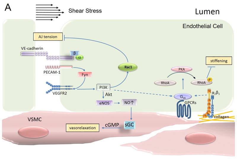
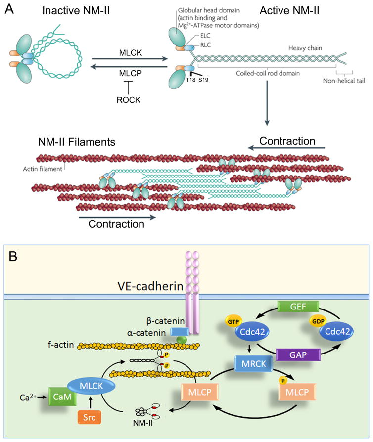
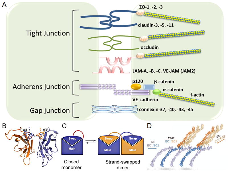
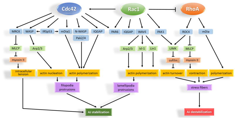
The monolayer of endothelial cells lining the vessel wall forms a semipermeable barrier (in all tissue except the relatively impermeable blood-brain and inner retinal barriers) that regulates tissue-fluid homeostasis, transport of nutrients, and migration of blood cells across the barrier. Permeability of the endothelial barrier is primarily regulated by a protein complex called adherens junctions. Adherens junctions are not static structures; they are continuously remodeled in response to mechanical and chemical cues in both physiological and pathological settings. Here, we discuss recent insights into the post-translational modifications of junctional proteins and signaling pathways regulating plasticity of adherens junctions and endothelial permeability. We also discuss in the context of what is already known and newly defined signaling pathways that mediate endothelial barrier leakiness (hyperpermeability) that are important in the pathogenesis of cardiovascular and lung diseases and vascular inflammation.

Keywords: adherens junctions; blood cells; endothelial cells; lung diseases; signal transduction.


Figures

Figure 1
Figure 1. Composition of inter-endothelial junctions
A) Schematic representation of inter-endothelial junctions comprised of tight junctions (TJs), adherens junctions (AJs), and gap junctions (GJs). TJs are mediated by adhesion proteins such as claudins, occludin, and junctional adhesion molecules (JAMs) whereas the zona occludin proteins (ZO-1, ZO-2 and ZO-3) connect adhesion molecules to the actin cytoskeleton. AJs are comprised of VE-cadherin and associated β- and p120-catenins. α-catenin binds β-catenin to connect AJs to the actin cytoskeleton. GJs are comprised of two connexin hexamers forming hemichannels. B) VE-cadherin mediates adhesion by trans-dimerization of tryptophan 2 and tryptophan 4 residues in a hydrophobic pocket of the opposing VE-cadherin molecule. Ribbon presentations of VE-cadherin trans-dimer. Adapted with permission from Brasch et al., Trends in Cell Biology, 2012. C) Strand swap dimerization occurs through the insertion of a tryptophan residue into the hydrophobic binding pocket of the opposing cadherin. Adapted with permission from Brasch et al., Trends in Cell Biology, 2012. D) Trans dimerization (between EC1 and EC1 of opposing cadherins) orients VE-cadherin molecules and facilitates cis interactions (between EC1 and EC2 of neighboring cadherins). Adapted with permission from Brasch et al., Trends in Cell Biology, 2012.
Figure 2
Figure 2. Role of acto-myosin apparatus in stabilizing AJs
A) Domain structure of non-muscle myosin II (NM-II). The NM-II consists of a globular head domain containing both actin-binding and motor domains, essential light chains (ELCs), regulatory light chains (RLCs), and heavy chains. NM-II possesses a head to tail interaction in the absence of phosphorylation. Phosphorylation of regulatory light chain at Thr18/Ser19 by myosin light chain kinase (MLCK) unfolds the molecule, enabling assembly of anti-parallel filaments through interactions between their rod domains. Activation of Rho-associated kinase (ROCK), which inhibits phosphatase activity of myosin light chain phosphatase (MLCP) in a phosphorylation-dependent manner, also favors RLC phosphorylation. NM-II filaments bind to actin filaments, which slide along each other, and cause a cell contraction. Adapted with permission from (Vicente-Manzanares et al., Nature Reviews Molecular Cell Biology, 2009). B) Proposed mechanism of regulation of NM-II activity at AJs in confluent endothelium. NM-II regulates attachment of the VE-cadherin adhesion complex to the actin cytoskeleton, thereby generating mechanical tension required for binding of α-catenin to both β-catenin and f-actin. NM-II phosphorylation is controlled by MLCK and MLCP activities. In the model, we propose that Src and Cdc42 pathways cooperate in regulating NM-II activity at AJs. Cdc42 facilitates activation of NM-II through myotonic dystrophy kinase-related Cdc42-binding kinase (MRCK)-dependent phosphorylation of MLCP whereas Src phosphorylates MLCK at sites of VE-cadherin adhesion. CaM = calmodulin; GEF = guanine nucleotide exchange factor; GAP = GTPase activating protein; GTP = guanosine triphosphate; GDP = guanosine diphosphate.
Figure 3
Figure 3. Mechanotransduction at AJs
The mechanosensory complex in endothelial cells is comprised of vascular endothelial (VE)-cadherin, platelet endothelial cell adhesion molecule (PECAM)-1 and vascular endothelial growth factor receptor (VEGFR)2. Mechanosensing of shear stress occurs through PECAM-1-dependent activation of Fyn, which in turn facilitates VEGFR2-mediated signaling in a ligand-independent manner and activates PI3K. PI3K activates both Rac1 and eNOS signaling pathways. Rac1 relieves tension at AJs whereas NO concomitantly promotes vasorelaxation of smooth muscle cells. PECAM-1-dependent sensing of shear stress also promotes α2β1 integrin signaling and consequently activation of PKA in atheroresistant regions. PKA phosphorylates RhoA and decreases RhoA-dependent cellular stiffness allowing the endothelial cell to align in the direction of blood flow. PI3K = phosphatidylinositol-4,5-bisphosphate 3-kinase; PKA = protein kinase A; VSMC = vascular smooth muscle cell; NO = nitric oxide; eNOS = endothelial nitric oxide synthase; cGMP = cyclic guanosine monophosphate; sGC = soluble guanylyl cyclase.
Figure 4
Figure 4. Role of specialized kinases and phosphatases in stabilizing AJs
A) Stable adherens junctions are characterized by low phosphorylation of VE-cadherin and associated catenin proteins. Protein tyrosine phosphatases DEP1, VE-PTP, PTPμ, SHP2, and PTP1B at AJs counteract the effect of tyrosine kinases (Src, Fen, Fyn, and Ableson) to stabilize the VE-cadherin-catenin complex. FAK also stabilizes VE-cadherin adhesion by inhibiting RhoA signaling through phosphorylation-dependent activation of p190RhoGAP. Ang1 = angiopoietin 1; VEGF = vascular endothelial growth factor ; VEGFR2 = vascular endothelial growth factor receptor 2; DEP1 = density enhanced phosphatase 1; VE-PTP = vascular endothelial protein tyrosine phosphatase; PTPμ = protein tyrosine phosphatase μ; SHP2 = Src homology phosphatase; PTP1B = protein tyrosine phosphatase 1 B; FAK = focal adhesion kinase; RhoGAP = Rho GTPase activating protein; RhoGEF = Rho guanine nucleotide exchange factor; GDP = guanosine diphosphate; GTP = guanosine triphosphate. B) Phosphorylation-dependent activation of kinases by VEGF, histamine, thrombin, PAF, and TNF-α leads to phosphorylation of VE-cadherin, β-catenin, and p120-catenin (residues are indicated) by distinct kinases. This results in destabilization of the VE-cadherin complex. Dissociation of p120-catenin due to phosphorylation of VE-cadherin at Y658 or p120-catenin at S879 exposes a VE-cadherin binding site for AP2 to facilitate VE-cadherin endocytosis via clathrin coated pits. Phosphorylation of β-catenin induces the uncoupling of VE-cadherin adhesion from the actin cytoskeleton. Activation of RhoA leads to phosphorylation of MLC, formation of stress fibers, and increased tension across VE-cadherin adhesion. TNF-α = tumor necrosis factor alpha; PAF = platelet-activating factor; AP2 = adaptor protein 2; MLC = myosin light chain; PKCα = protein kinase C alpha.
Figure 5
Figure 5. Regulation of RhoGTPase activity
A) Schematic representation of general domain structure for RhoGTPases. The 5 G-box motifs (green) represent nucleotide binding motifs whereas the switch I and Switch II are the region of GDP/GTP exchange. C-terminus (red) undergoes post translational modification required for modulating the membrane-targeting of RhoGTPases. B) Conformational changes within Switch I and II regions upon GTP hydrolysis and exchange. The closed GTP-bound conformation has a higher affinity for GAP binding. Cleavage of hydrolyzed phosphate by GAPs put the switch regions into a relaxed, open conformation. The open GDP-bound conformation has a high affinity for GEF binding. GAP = GTPase activating protein; GEF = guanine nucleotide exchange factor; GDP = guanosine diphosphate; GTP = guanosine triphosphate; GNBP = guanine nucleotide binding protein. Adapted with permission from Vetter and Wittinghofer, Science, 2001. C) Regulation of RhoGTPase cycle. In the GDP-bound state, RhoGTPases are prevented from interacting with downstream effectors. Release of GDP is facilitated by GEFs allowing exchange for GTP. GAPs catalyze the hydrolysis of GTP resulting in inactivation of the GTPase. GDIs prevent GTP exchange by binding to the GDP bound state. GDI = guanosine nucleotide dissociation inhibitor. Adapted with permission from Etienne-Manneville and Hall; Nature, 2002.
Figure 6
Figure 6. RhoA, Rac1 and Cdc42 regulation of endothelial AJs
Rac1 and Cdc42 promote organization of the actin cytoskeleton into lamellipodia and filopodia protrusions resulting in re-annealing and stabilization of AJs. Regardless of differential effect on actin organization, Rac1 and Cdc42 share common downstream effectors such as PAR6 and IQGAP1. These effectors serve as scaffolds by recruiting active Rac1 and Cdc42 to AJs. Cdc42 can also generate low grade tension at AJs through activation of non-muscle myosin II. In contrast to Rac1 and Cdc42, RhoA activity is basally suppressed at AJs. Activation of RhoA is associated with formation of stress fibers, increased intracellular tension, and destabilization of AJs. MRCK = myotonic dystrophy kinase-related Cdc42-binding kinase; WASP = Wiskott-Aldrich Syndrome protein; IRSp53 = insulin receptor tyrosine kinase substrate p53; mDia = mammalian Diaphanous; Pak = p21 activated kinase; IQGAP = IQ motif containing GTPase activating protein; PAR6 = partitioning defective protein 6; WAVE = Wasp family verproline-homologue; MLCP = myosin light chain phosphatase; Arp2/3 = Actin-related proteins 2 and 3; LIMK = LIM (Lin1, Isl-1, & Mec-3) kinase; Isl-1 = Insulin gene enhancer protein; Lin1 = CD2 cytoplasmic tail binding protein 2.

Comment in

Similar articles

Cited by

References

    1. Pappenheimer JR, Renkin EM, Borrero LM. Filtration, diffusion and molecular sieving through peripheral capillary membranes; a contribution to the pore theory of capillary permeability. The American journal of physiology. 1951;167:13–46. - PubMed
    1. Del Vecchio PJ, Siflinger-Birnboim A, Shepard JM, Bizios R, Cooper JA, Malik AB. Endothelial monolayer permeability to macromolecules. Federation proceedings. 1987;46:2511–2515. - PubMed
    1. Siflinger-Birnboim A, Del Vecchio PJ, Cooper JA, Blumenstock FA, Shepard JM, Malik AB. Molecular sieving characteristics of the cultured endothelial monolayer. J Cell Physiol. 1987;132:111–117. - PubMed
    1. Schneeberger EE. Circulating proteins and macromolecular transport across continuous, nonfenestrated endothelium. Annals of the New York Academy of Sciences. 1982;401:25–37. - PubMed
    1. Weisberg HF. Osmotic pressure of the serum proteins. Annals of clinical and laboratory science. 1978;8:155–164. - PubMed

MeSH terms

LinkOut - more resources