

doi: 10.1038/srep39846.
Isolation of cell type-specific apoptotic bodies by fluorescence-activated cell sorting
Affiliations
- PMID: 28057919
- PMCID: PMC5216387
- DOI: 10.1038/srep39846
Item in Clipboard
Isolation of cell type-specific apoptotic bodies by fluorescence-activated cell sorting
Sci Rep.
.
Abstract
Apoptotic bodies (ApoBDs) are membrane-bound extracellular vesicles that can mediate intercellular communication in physiological and pathological settings. By combining recently developed analytical strategies with fluorescence-activated cell sorting (FACS), we have developed a method that enables the isolation of ApoBDs from cultured cells to 99% purity. In addition, this approach also enables the identification and isolation of cell type-specific ApoBDs from tissue, bodily fluid and blood-derived samples.
Figures
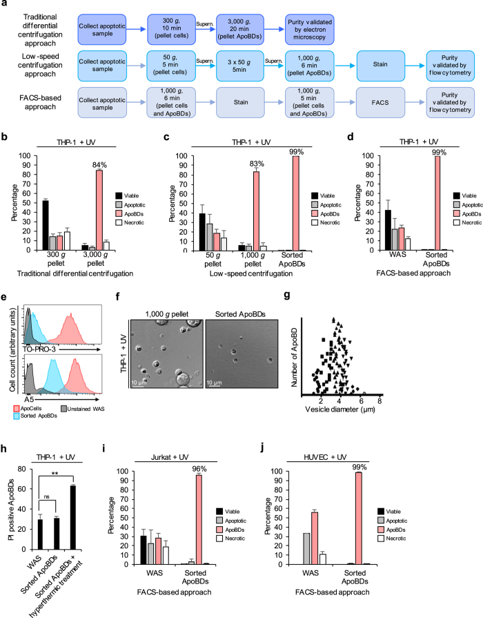
(a) Schematic diagram of ApoBD purification steps for two pre-existing centrifugation approaches, and the newly developed FACS-based approach (supern = supernatant). (b–d) Flow cytometry analysis of the purity of THP-1 monocyte-derived ApoBDs isolated by (b) traditional differential centrifugation approach, (c) low-speed centrifugation approach, and (d) FACS-based approach (n = 3). (e) TO-PRO-3 and A5 staining of THP-1 apoptotic cells and sorted ApoBDs compared to unstained WAS, data generated from (d). (f) Representative DIC microscopy showing ApoBDs purified by low-speed centrifugation approach alone or in combination with FACS-based approach. (g) Diameter (μm) of sorted ApoBDs generated from THP-1 monocytes (representative of 1 independent experiment, n = 3). (h) PI uptake by ApoBDs from THP-1 WAS, sorted ApoBDs and sorted ApoBDs exposed to hyperthermic treatment (n = 3). (i,j) Purity of ApoBDs isolated from apoptotic Jurkat T cells (i) and HUVEC (j) WAS by FACS-based approach (n = 3). Error bars represent s.e.m. (n = independent experiment). Statistical significant differences determined by two-tailed t-test. ns, not significant. **P < 0.01.
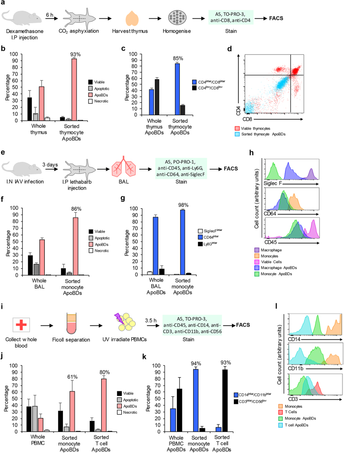
(a) Schematic diagram of the dexamethasone mouse model used to generate and purify thymocyte-derived ApoBDs. (b) Purity of sorted thymocyte ApoBDs compared to whole thymus sample from dexamethasone-treated mice (n = 3). (c) Percentage of ApoBDs expressing intermediate CD4/CD8 and low CD4/CD8 from whole thymus or sorted ApoBD samples (n = 3). (d) CD4 and CD8 staining of viable thymocytes and sorted thymocyte ApoBDs. (e) Schematic diagram of the IAV mouse model used to generate and isolate BAL-derived monocyte ApoBDs. (f) Purity of sorted monocyte ApoBDs compared to whole BAL sample from IAV-infected mice (n = 3). (g) Percentage of ApoBDs expressing intermediate levels of SiglecF, CD64 or Ly6G from whole BAL or sorted ApoBD samples (n = 3). (h) Levels of SiglecF, CD64 and CD45 staining on macrophages, macrophage-derived ApoBDs, monocytes, monocyte-derived ApoBDs and total viable cells. (i) Schematic diagram of the PBMC model used to generate and purify human monocyte and T cell-derived ApoBDs. (j) Purity of sorted monocyte and T cell ApoBDs isolated from apoptotic PBMC samples (n = 3). (k) Percentage of ApoBDs expressing intermediate CD14/CD11b and intermediate CD3/low CD56 from whole PBMC, sorted monocyte and sorted T cell ApoBD samples (n = 3). (l) Levels of CD14, CD11b and CD3 expression on monocytes, monocyte ApoBDs, T cells and T cell ApoBDs. Error bars represent s.e.m. (n = independent experiment).
References
-
- Schiller M. et al. Autoantigens are translocated into small apoptotic bodies during early stages of apoptosis. Cell Death Differ 15, 183–91 (2008). - PubMed
-
- Cocca B. A., Cline A. M. & Radic M. Z. Blebs and apoptotic bodies are B cell autoantigens. J Immunol 169, 159–66 (2002). - PubMed
-
- Tran H. B. et al. Subcellular redistribution of la/SSB autoantigen during physiologic apoptosis in the fetal mouse heart and conduction system: a clue to the pathogenesis of congenital heart block. Arthritis Rheum 46, 202–8 (2002). - PubMed
Publication types
MeSH terms
LinkOut - more resources
Full Text Sources
Other Literature Sources
