

A simple and rapid flow cytometry-based assay to identify a competent embryo prior to embryo transfer
- PMID: 28057937
- PMCID: PMC5216337
- DOI: 10.1038/srep39927
A simple and rapid flow cytometry-based assay to identify a competent embryo prior to embryo transfer
Abstract
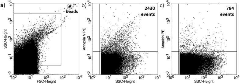
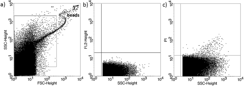
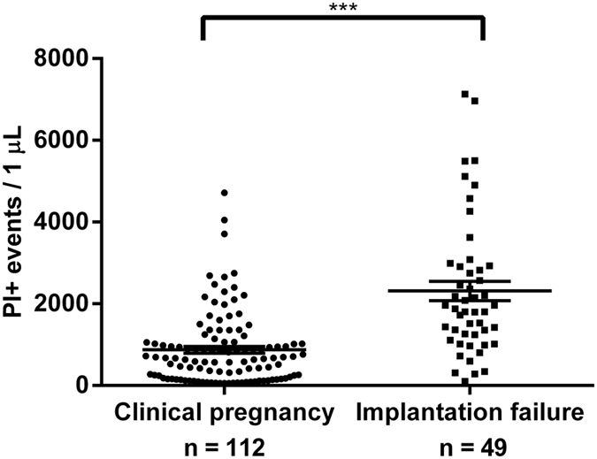
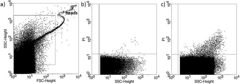
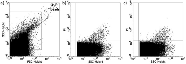
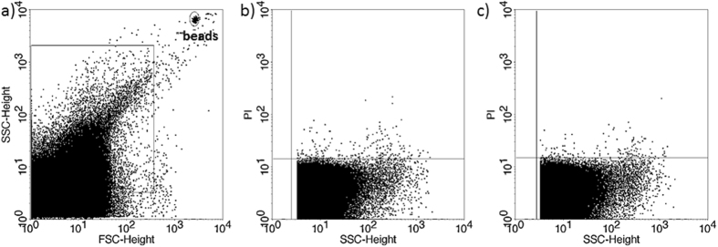
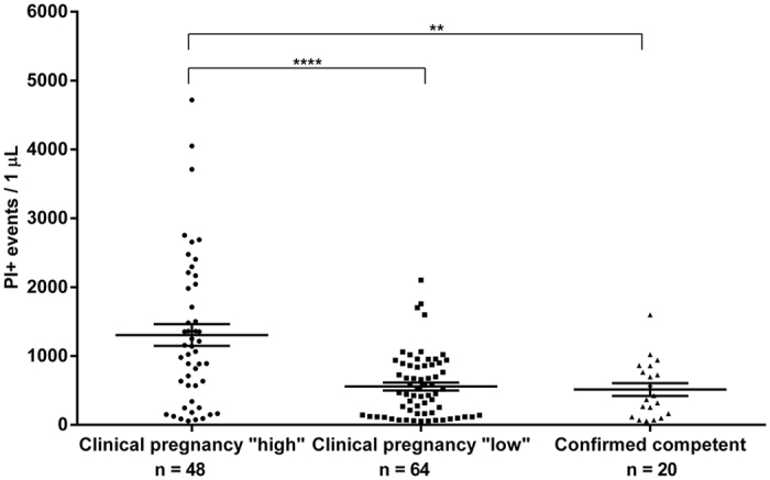
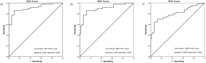
Multiple pregnancy is a risk for prematurity and preterm birth. The goal of assisted reproduction is to achieve a single pregnancy, by transferring a single embryo. This requires improved methods to identify the competent embryo. Here, we describe such a test, based on flow cytometric determination of the nucleic acid (PI+) containing extracellular vesicle (EV) count in day 5 embryo culture media. 88 women undergoing IVF were included in the study. More than 1 embryos were transferred to most patients. In 58 women, the transfer resulted in clinical pregnancy, whereas in 30 women in implantation failure. In 112 culture media of embryos from the "clinical pregnancy" group, the number of PI+ EVs was significantly lower than in those of 49 embryos, from the "implantation failure" group. In 14 women, transfer of a single embryo resulted in a singleton pregnancy, or, transfer of two embryos in twin pregnancy. The culture media of 19 out of the 20 "confirmed competent" embryos contained a lower level of PI+ EVs than the cut off level, suggesting that the competent embryo can indeed be identified by low PI+ EV counts. We developed a noninvasive, simple, inexpensive, quick test, which identifies the embryos that are most likely to implant.
Figures
Similar articles
-
Noninvasive metabolomic profiling of embryo culture media using proton nuclear magnetic resonance correlates with reproductive potential of embryos in women undergoing in vitro fertilization.Fertil Steril. 2008 Dec;90(6):2183-9. doi: 10.1016/j.fertnstert.2008.07.1739. Epub 2008 Oct 8. Fertil Steril. 2008. PMID: 18842260
-
Extracellular vesicles secreted during blastulation show viability of bovine embryos.Reproduction. 2019 Dec;158(6):477-492. doi: 10.1530/REP-19-0233. Reproduction. 2019. PMID: 31600718
-
Guidelines for the number of embryos to transfer following in vitro fertilization No. 182, September 2006.Int J Gynaecol Obstet. 2008 Aug;102(2):203-16. doi: 10.1016/j.ijgo.2008.01.007. Int J Gynaecol Obstet. 2008. PMID: 18773532 Review.
-
Soluble human leukocyte antigen G expression in phase I culture media at 46 hours after fertilization predicts pregnancy and implantation from day 3 embryo transfer.Fertil Steril. 2005 May;83(5):1410-3. doi: 10.1016/j.fertnstert.2004.11.061. Fertil Steril. 2005. PMID: 15866577
-
Temperature of embryo culture for assisted reproduction.Cochrane Database Syst Rev. 2019 Sep 17;9(9):CD012192. doi: 10.1002/14651858.CD012192.pub2. Cochrane Database Syst Rev. 2019. PMID: 31529804 Free PMC article.
Cited by
-
PIBF+ extracellular vesicles from mouse embryos affect IL-10 production by CD8+ cells.Sci Rep. 2018 Mar 16;8(1):4662. doi: 10.1038/s41598-018-23112-z. Sci Rep. 2018. PMID: 29549351 Free PMC article.
-
Preimplantation genetic testing in the current era, a review.Arch Gynecol Obstet. 2024 May;309(5):1787-1799. doi: 10.1007/s00404-024-07370-z. Epub 2024 Feb 20. Arch Gynecol Obstet. 2024. PMID: 38376520 Review.
-
Noninvasive Biomarkers of Human Embryo Developmental Potential.Int J Mol Sci. 2025 May 21;26(10):4928. doi: 10.3390/ijms26104928. Int J Mol Sci. 2025. PMID: 40430065 Free PMC article. Review.
-
Long-Term Effects of ART on the Health of the Offspring.Int J Mol Sci. 2023 Sep 1;24(17):13564. doi: 10.3390/ijms241713564. Int J Mol Sci. 2023. PMID: 37686370 Free PMC article. Review.
-
MicroRNAs from Extracellular Vesicles Secreted by Bovine Embryos as Early Biomarkers of Developmental Competence.Int J Mol Sci. 2020 Nov 24;21(23):8888. doi: 10.3390/ijms21238888. Int J Mol Sci. 2020. PMID: 33255183 Free PMC article.
References
-
- Munné S., Alikani M., Tomkin G., Grifo J. & Cohen J. Embryo morphology, developmental rates and maternal age are correlated with chromosome abnormalities. Fertil Steril ; 64, 382–391 (1995). - PubMed
-
- Magli M. C., Gianaroli L. & Ferrareti A. P. Chromosomal abnormalities in embryos. Mol. Cell. Endocrinol. 183(suppl. 1), S29–S34 (2001). - PubMed
-
- Márquez C., Sandalinas M., Bahçe M., Alikani M. & Munné S. Chromosome abnormalities in 1255 cleavage-stage human embryos. Reprod. BioMed. Online 1, 17–26 (2000). - PubMed
-
- Bielanska M., Tan S. L. & Ao A. High rate of mixoploidy among human blastocysts cultured in vitro. Fertil. Steril. 78, 1248–53 (2002). - PubMed
-
- Rai R. & Regan L. Recurrent miscarriage. Lancet ; 368, 601–611 (2006). - PubMed
Publication types
MeSH terms
Substances
LinkOut - more resources
Full Text Sources
Other Literature Sources
Research Materials
Miscellaneous
