Comparative Study
. 2017 Oct;31(10):2122-2131.
doi: 10.1038/leu.2017.1. Epub 2017 Jan 6.

Activating JAK2 mutants reveal cytokine receptor coupling differences that impact outcomes in myeloproliferative neoplasm

Affiliations
Comparative Study

Activating JAK2 mutants reveal cytokine receptor coupling differences that impact outcomes in myeloproliferative neoplasm

H Yao et al. Leukemia. 2017 Oct.

Abstract

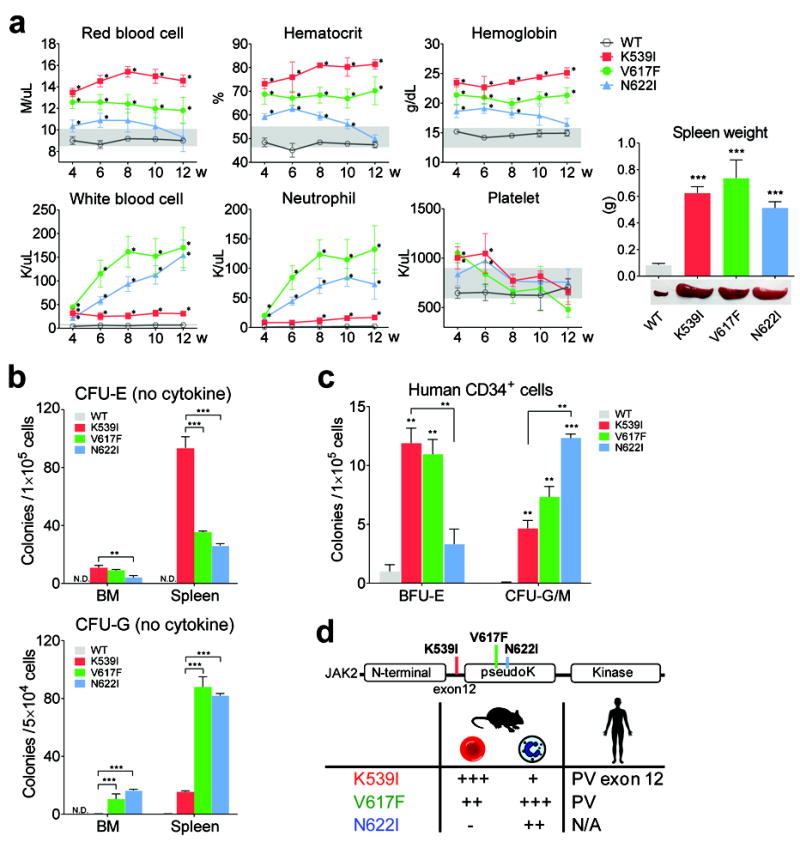
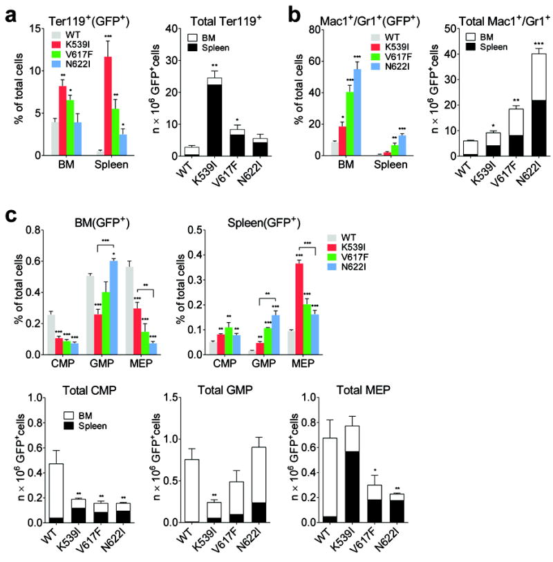
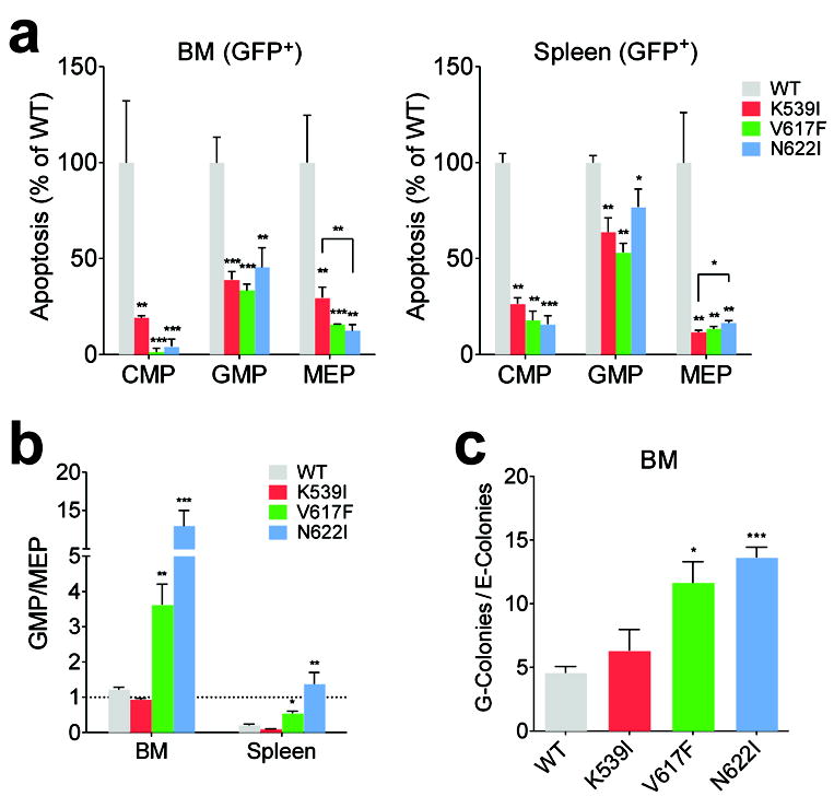
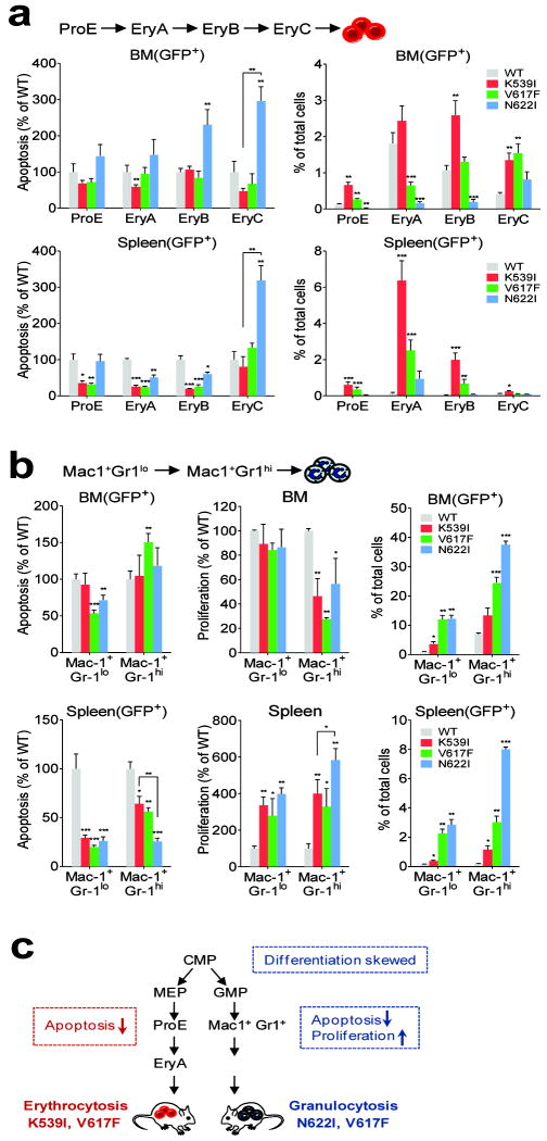
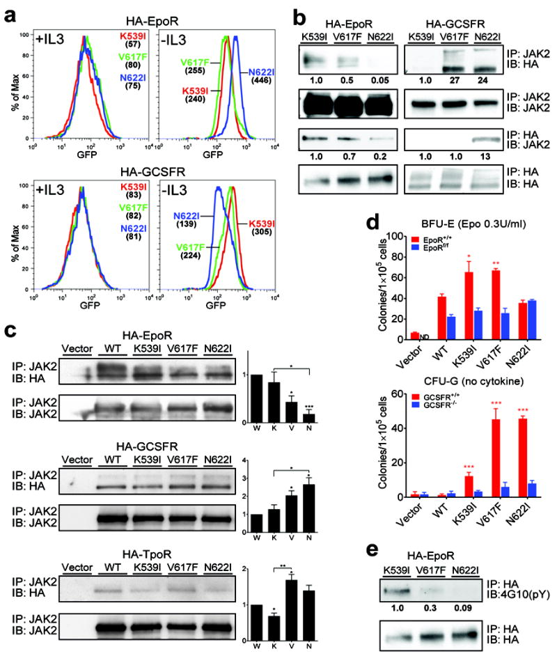
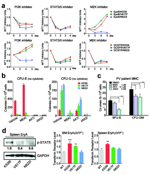
Janus tyrosine kinase 2 (JAK2) mediates downstream signaling of cytokine receptors in all hematological lineages, yet constitutively active JAK2 mutants are able to drive selective expansion of particular lineage(s) in myeloproliferative neoplasm (MPN). The molecular basis of lineage specificity is unclear. Here, we show that three activating JAK2 mutants with similar kinase activities in vitro elicit distinctive MPN phenotypes in mice by differentially expanding erythroid vs granulocytic precursors. Molecularly, this reflects the differential binding of JAK2 mutants to cytokine receptors EpoR and GCSFR in the erythroid vs granulocytic lineage and the creation of unique receptor/JAK2 complexes that generate qualitatively distinct downstream signals. Our results demonstrate that activating JAK2 mutants can differentially couple to selective cytokine receptors and change the signaling repertoire, revealing the molecular basis for phenotypic differences elicited by JAK2 (V617F) or mutations in exon 12. On the basis of these findings, receptor-JAK2 interactions could represent new targets of lineage-specific therapeutic approaches against MPN, which may be applicable to other cancers with aberrant JAK-STAT signaling.


Conflict of interest statement

CONFLICT OF INTEREST

The authors declare no conflict of interest.

Figures

Fig. 1
Fig. 1
Activating JAK2 mutants confer distinct hematological phenotypes in mice. (a) Hematological parameters of mice assessed at indicated times post transplantation. Grey zones indicate normal range. w: weeks post transplantation. *p<0.01 (vs. WT). Mice expressing JAK2 mutants exhibited splenomegaly at 12 weeks post transplantation. (b) Cells from JAK2 mutant mice but not WT mice generated cytokine-independent erythroid and granulocytic colonies. *p<0.05, **p<0.01, ***p<0.001. n=3 in each group.ND: not detected. (c) Expression of JAK2 mutants in human CD34+ cells differentially affects erythroid vs. granulocytic lineages. Normal CD34+ cells were transduced with lentiviruses expressing WT or mutant JAK2 and plated in methylcellulose media with puromycin to select for transduced cells. Epo-independent BFU-E colonies and cytokine-independent CFU-G/M colonies, which include CFU-G, CFU-M and CFU-GM colonies are shown. Colonies were enumerated on day 14. n=3 in each group. *p<0.05, **p<0.01, ***p<0.001 (vs. WT unless specified). (d) Summary of blood phenotypes. The degree of erythrocytosis and granulocytosis are indicated by plus signs.Results represent at least three independent experiments.
Fig. 2
Fig. 2
Erythroid and granulocytic precursors are differentially expanded in JAK2 mutant mice. (a-b)Frequency and total number of Ter119+ erythroid (a) and Mac1+/Gr1+ granulocytic (b) precursors were increased in JAK2 mutant mice. Because hind limbs BM represents approximately 25% of total bone marrow , total number of precursors per animal was calculated as the sum of the number from spleen and four fold the number from hind limbs BM. (c) Frequency and total number of BM and splenic CMP, MEP, and GMP progenitors from mice expressing JAK2 mutants or WT mice. In all panels, GFP+ indicates that GFP+ cells are gated for analyses. n=3 in each group. *p<0.05, **p<0.01, ***p<0.001 (vs. WT unless specified). Results represent at least three independent experiments.
Fig. 3
Fig. 3
Survival and differentiation analyses of myeloid progenitors from JAK2 mutant mice. (a) Annexin V-binding analyses in CMP, MEP, and GMP progenitors normalized to WT controls.Apoptotic cells (GFP+AnnexinV+7AAD- plus GFP+AnnexinV+7AAD+) were quantified. (b) GFP+ GMP to MEP ratio is significantly higher in JAK2(V617F) and JAK2(N622I) mice compared to WT mice. (c) Sorted GFP+ CMP from JAK2(V617F) and JAK2(N622I) mice generated more granulocytic and macrophage colonies (G-colonies) than erythroid and megakaryocytic colonies (E-colonies) compared to WT mice. n=3 in each group, *p<0.05, **p<0.01, ***p<0.001 (vs. WT unless specified). Results represent at least three independent experiments.
Fig. 4
Fig. 4
Survival and proliferation analyses of erythroid and myeloid precursor subsets from JAK2 mutant mice. (a) Quantitative analyses of Annexin V-binding in erythroblast subsets normalized to WT controls. Early apoptotic cells (GFP+AnnexinV+7AAD-), and frequency of BM and splenic erythroblast subsets were quantified. (b)Quantitative analyses of Annexin V-binding and BrdU incorporation in granulocytic precursor subsets normalized to WT controls. Early apoptotic cells (GFP+AnnexinV+7AAD-) were shown. Frequency of BM and splenic granulocytic precursor subsets were quantified. (c) Summary of cellular effects of JAK2 mutants.n=3 in each group. *p<0.05, **p<0.01, ***p<0.001 (vs. WT unless specified). Results represent two independent experiments.
Fig. 5
Fig. 5
JAK2 mutants interact with cytokine receptors in different ways. (a) The amount of co-expressed receptors necessary for each JAK2 mutant to transform BaF3 is different. BaF3 cells stably expressing WT or mutant JAK2 with HA-tagged EpoR or GCSFR (bicistronically co-expressed with GFP) were selected in media without IL3. Median GFP fluorescence intensities (numbers in parentheses) indicate receptor expression level before and after IL3 withdrawal. (b) JAK2 mutants bind to receptors with different affinities. Co-immunoprecipitation analyses of JAK2 mutants and HA-tagged receptors in BaF3 cells using either JAK2 or HA antibodies.Immunoblot band intensity was quantified by ImageJ software and normalized to JAK2(K539I). (c) JAK2 mutants differentially bind to receptors in JAK2-deficient γ2A cells. JAK2 mutants were transiently expressed in γ2A cells in which HA-tagged EpoR, GCSFR, or TpoR were stably expressed. Interaction was examined by co-immunoprecipitation with JAK2 antibodies. Immunoblot band intensity was quantified by ImageJ software and normalized to loading control and wild-type JAK2. W:WT, K: K539I, V: V617F, N: N622I. (d) Deletion of EpoR or GCSFR impairs hypersensitive erythroid colonies and cytokine-independent granulocytic colonies produced by JAK2 mutants. Bone marrow cells from wild-type or EpoRflox/flox mice, transduced to express JAK2 mutants and GFPCre, and bone marrow cells from wild-type or GCSFR-/- mice, transduced to express JAK2 mutants, were plated in methylcellulose media (M3234) with 0.3U/ml Epo (for BFU-E) or with no cytokine (for CFU-G). Colonies were enumerated on day 5 and 7. (e) EpoR is tyrosine phosphorylated in cells expressing JAK2(K539I) and JAK2(V617F) but not JAK2(N622I). HA-EpoR immunoprecipitated via HA agarose was probed with anti-phosphotyrosine antibody 4G10. *p<0.05, **p<0.01, ***p<0.001 (vs. WT unless specified). ND: not detected. IP: immunoprecipitation. IB: immunoblot. Results represent at least three independent experiments.
Fig. 6
Fig. 6
JAK2 mutants elicit distinct downstream signaling from receptor-JAK2 complex. (a) Sensitivity of BaF3 cells expressing JAK2 mutants with EpoR (top panels) or GCSFR (bottom panels) to inhibitors of PI3K (LY294002), STAT3/5 (pimozide) and MEK (U0126). (b) Cytokine-independent erythroid colonies and granulocytic colonies from JAK2 mutant mice grown in the presence of inhibitors to MEK (AZD6244), AKT (MK2206) or JAK2 (AZD1480) were enumerated.n=3 in each group. (c) Effects of inhibitors on the formation of Epo-independent BFU-E and cytokine-independent granulocytic colonies from JAK2(V617F)-positive PV patient mononuclear cells (MNC). Colonies were enumerated on day 14. n=3 in each group. (d) Levels of phospho-STAT5 and its target gene transferrin receptor are highest in sorted EryA cells expressing JAK2(K539I) among the three mutants. Immunoblot band intensity was quantified by ImageJ software and normalized to JAK2(K539I). *p<0.05, **p<0.01, ***p<0.001 (vs. vehicle (DMSO) in (b-c) or WT (d)). ND: not detected. Results represent at least three independent experiments.

Similar articles

Cited by

References

    1. Thomas SJ, Snowden JA, Zeidler MP, Danson SJ. The role of JAK/STAT signalling in the pathogenesis, prognosis and treatment of solid tumours. Br J Cancer. 2015 Jul 28;113(3):365–71. - PMC - PubMed
    1. Vainchenker W, Constantinescu SN. JAK/STAT signaling in hematological malignancies. Oncogene. 2013 May 23;32(21):2601–13. - PubMed
    1. Villarino AV, Kanno Y, Ferdinand JR, O’Shea JJ. Mechanisms of Jak/STAT signaling in immunity and disease. J Immunol. 2015 Jan 1;194(1):21–7. - PMC - PubMed
    1. Stark GR, Darnell JE., Jr The JAK-STAT pathway at twenty. Immunity. 2012 Apr 20;36(4):503–14. - PMC - PubMed
    1. Levine RL. JAK-mutant myeloproliferative neoplasms. Curr Top Microbiol Immunol. 2012 Aug 7;355:119–33. - PubMed

Publication types

MeSH terms

Substances

LinkOut - more resources