Review
. 2016 Nov-Dec;49(6):389-396.
doi: 10.1590/0100-3984.2015.0076.

Ultrasound findings of the physiological changes and most common breast diseases during pregnancy and lactation

Affiliations
Review

Ultrasound findings of the physiological changes and most common breast diseases during pregnancy and lactation

Antônio Arildo Reginaldo de Holanda et al. Radiol Bras. 2016 Nov-Dec.

Abstract

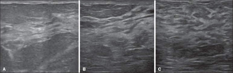
Because of the physiological changes that occur during pregnancy and lactation, diagnostic ultrasound of the breast during these periods is a challenge for physicians. Therefore, a comprehensive understanding of imaging, anatomy, and physiology of the breast is important to effectively diagnosing diseases that can arise in women who are pregnancy or lactating. The aim of this article was to review the physiological changes that occur in the breasts during pregnancy and lactation, as well as to describe the main features of the breast diseases that occur most frequently during these periods.

O diagnóstico ultrassonográfico das mamas durante a gravidez e lactação representa um desafio para o médico, em função das alterações fisiológicas próprias destes períodos. Para tanto, é essencial uma compreensão das imagens, da anatomia e da fisiologia mamárias para diagnosticar mais eficazmente doenças concomitantes. O presente artigo teve como objetivo fazer uma revisão das alterações fisiológicas que ocorrem nas mamas durante a gravidez e lactação, bem como relatar as principais características ultrassonográficas das doenças mamárias mais frequentes nestes períodos.

Keywords: Breast; Lactation; Pregnancy; Ultrasonography.


Figures

Figure 1
Figure 1
A: Breast in the first trimester of pregnancy: predominantly hypoechoic breast parenchyma, showing dilatation of the milk ducts. B: Breast in the second trimester of pregnancy: breast parenchyma showing greater echogenicity and more pronounced ductal dilatation than in the first trimester. C: Breast in the third trimester of pregnancy: breast parenchyma showing considerably greater echogenicity and extremely more pronounced ductal dilatation in comparison with the second trimester.
Figure 2
Figure 2
Breast during lactation. Diffusely hyperechoic breast parenchyma, with ductal dilatation due to the accumulation of milk.
Figure 3
Figure 3
Puerperal mastitis. Amorphous areas of variable echogenicity, predominantly hypoechoic and heterogeneous, diffusely distributed throughout the breast parenchyma, with poorly defined borders.
Figure 4
Figure 4
Abscess. A: Amorphous, unilocular complex lesion, with ill-defined borders, parallel to the skin, heterogeneous, with variable echogenicity, that is predominantly hypoechoic, with discrete posterior acoustic enhancement, corresponding to an abscess with a fluctuant area. B: Ovoid lesion, with well-defined borders, parallel to the skin, hypoechoic, homogeneous, featuring discrete posterior acoustic enhancement, corresponding to abscess formation.
Figure 5
Figure 5
Benign reactive lymph node. Ovoid nodule, parallel to the skin, with well-defined, hypoechoic borders, and a hyperechoic halo, corresponding to a benign lymph node.
Figure 6
Figure 6
Granulomatous mastitis. Amorphous formation, not parallel to the skin, with ill-defined, hypoechoic borders.
Figure 7
Figure 7
Galactocele. A: Lesion, parallel to the skin, with well-defined borders, showing anechoic (cystic) and echogenic (solid) components, with discrete posterior acoustic enhancement and well-defined borders. B: Predominantly hypoechoic lesion, parallel to the skin, with welldefined borders, peripheral areas of hyperechogenicity, and posterior acoustic enhancement.
Figure 8
Figure 8
Lactating adenoma. Ovoid nodule, parallel to the skin, with a heterogeneous, hypoechoic pattern and well-defined borders.
Figure 9
Figure 9
Fibroadenoma. Ovoid nodule, parallel to the skin, with a homogeneous, hypoechoic pattern, well-defined borders, and discrete posterior acoustic enhancement.
Figure 10
Figure 10
Pregnancy-associated breast cancer. A: Amorphous formation, parallel to the skin, with variable echogenicity (predominantly hypoechoic), a heterogeneous texture, discrete acoustic enhancement and poorly defined borders. B: Irregular, hypoechoic, heterogeneous nodule, parallel to the skin, featuring discrete acoustic shadowing and poorly defined borders.

References

    1. Yu JH, Kim MJ, Cho H, et al. Breast diseases during pregnancy and lactation. Obstet Gynecol Sci. 2013;56:143–159. - PMC - PubMed
    1. Joshi S, Dialani V, Marotti J, et al. Breast disease in the pregnant and lactating patient: radiological-pathological correlation. Insights Imaging. 2013;4:527–538. - PMC - PubMed
    1. Boisserie-Lacroix M, Dos Santos E, Belléannée G, et al. La femme enceinte: difficultés diagnostiques. Imagerie de la Femme. 2004;14:145–152.
    1. Canoy JM, Mitchell GS, Unold D, et al. A radiologic review of common breast disorders in pregnancy and the perinatal period. Semin Ultrasound CT MR. 2012;33:78–85. - PubMed
    1. Svensson WE. A review of the current status of breast ultrasound. Eur J Ultrasound. 1997;6:77–101.