. 2016 Dec 21;22(47):10353-10363.
doi: 10.3748/wjg.v22.i47.10353.

Vitamin D differentially regulates Salmonella-induced intestine epithelial autophagy and interleukin-1β expression

Affiliations

Vitamin D differentially regulates Salmonella-induced intestine epithelial autophagy and interleukin-1β expression

Fu-Chen Huang. World J Gastroenterol. .

Abstract

Aim: To investigate the effects of active vitamin D3 on autophagy and interleukin (IL)-1β expression in Salmonella-infected intestinal epithelial cells (IECs).

Methods: Caco-2 cells, NOD2 siRNA-, Atg16L1 siRNA- or vitamin D receptor (VDR) siRNA-transfected Caco-2 cells were pretreated with 1,25-dihydroxyvitamin D3 (1,25D3), and then infected by wild-type S. typhimurium strain SL1344. The conversion of LC3-I to LC3-II was detected by Western blot analysis and LC3+ autophagosome was analyzed by immunofluorescence. Caco-2 cells or VDR siRNA-transfected cells were pretreated with 1,25D3, and then infected by SL1344. Membrane protein and total RNA were analyzed by Western blot and RT-PCR for VDR and Atg16L1 protein and mRNA expression, respectively. Atg16L1 siRNA-transfected Caco-2 cells were pretreated by 1,25D3 and then infected with SL1344. Total RNA was analyzed by RT-PCR for IL-1β mRNA expression.

Results: The active form of vitamin D, 1,25D3, showed enhanced VDR-mediated Atg16L1 mRNA expression, membranous Atg16L1 protein expression leading to enhanced autophagic LC3II protein expression and LC3 punctae in Salmonella-infected Caco-2 cells which was counteracted by Atg16L1 and VDR siRNA, but Atg16L1 mediated suppression of IL-1β expression. Thus, active vitamin D may enhance autophagy but suppress inflammatory IL-1β expression in Salmonella-infected IECs.

Conclusion: Active vitamin D might enhance autophagic clearance of Salmonella infection, while modulation of inflammatory responses prevents the host from detrimental effects of overwhelming inflammation.

Keywords: Atg16L1; Autophagy; Interleukin-1β; Intestinal epithelia; Salmonella; Vitamin D.


Conflict of interest statement

Conflict-of-interest statement: The author declares that there are no financial or commercial conflicts of interest.

Figures
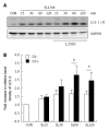
Figure 1
Figure 1
Effect of 1,25D3 on autophagy expression in Salmonella-infected Caco-2 cells. Caco-2 cells were untreated (CON) or treated with 1,25D3 (D3) and then infected by S. typhimurium wild-type strain SL1344 (SL). Immunoblots were performed on whole cell lysates with antibody to detect autophagy LC3-I/II protein expression. The results shown are representative of three separate experiments. GAPDH was detected for normalization of cytosolic protein. Representative immunoblots (A) and densitometric quantification of immunoreactive bands (B) are shown. The relative band intensities of LC3-II in untreated (white) and treated (black) Caco-2 cells were quantified as fold-increases compared with the control cells.
Figure 2
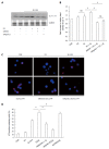
Figure 2
Involvement of NOD2 and Atg16L1 in 1,25D3-enhanced autophagy expression in Salmonella-infected Caco-2 cells. Caco-2 cells were transfected with control siRNA, NOD2 siRNA or Atg16L1 siRNA (siCON: non-target control siRNA; siNOD2: siRNA to NOD2; siAtg16L1: siRNA to Atg16L1) for 48 h. The transfected cells were left uninfected (CON) or infected by wild-type S. typhimurium strain SL1344 (SL) in the absence or presence of 1,25D3 (D3). Knockdown of NOD2[21] and Atg16L1[18] was confirmed previously by Western blot. The transfected cells were pretreated with 1,25D3 prior to wild-type S. typhimurium strain SL1344 infection for 1 h. Immunoblotting was performed on whole cell lysates with antibody to detect autophagy LC3-I/II protein expression. The results shown are representative of three separate experiments. GAPDH was detected for normalization of cytosolic protein. Representative immunoblots (A) and densitometric quantification of immunoreactive bands (B) are shown. The relative band intensities of LC3-II in Caco-2 cells are quantified as fold-increases compared with the control cells. The cells were fixed, permeabilized and stained with anti-LC3 (red), and LC3 puncta formation was detected by a confocal microscope; representative images of LC3 punctae are depicted. Scale bar = 10 μm (C); The percentage of cells showing accumulation of LC3 punctae is reported (D). Data are mean ± SEM of three independent experiments. aP < 0.05.
Figure 3
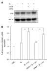
Figure 3
Effect of 1,25D3 on Atg16L1 mRNA expressions in Salmonella-infected Caco-2 cells and involvement of vitamin D receptor in the effect. Caco-2 cells were transfected with control siRNA and vitamin D receptor (VDR) siRNA (siRNA: non-target control siRNA; siVDR: siRNA to VDR) for 48 h. The transfected cells were left uninfected (CON) or infected by wild-type S. typhimurium strain SL1344 in the absence or presence of 1,25D3. Western blots probed with antibodies against VDR and GAPDH confirmed knockdown of VDR (A). Total RNA was prepared after infection and analyzed by real-time quantitative PCR to estimate amounts of Atg16L1 transcript. The amount of Atg16L1 mRNA produced and normalized to the corresponding amount of GAPDH transcript is shown as the fold-increase over uninfected control cells (B). Results are presented as mean ± SEM for at least three determinations from independent experiments. aP < 0.05.
Figure 4
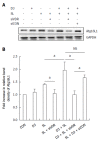
Figure 4
Effect of 1,25D3 on the membranous Atg16L1 protein expression in Salmonella-infected Caco-2 cells via vitamin D receptor. Caco-2 cells were transfected with control siRNA and vitamin D receptor (VDR) siRNA (siCON: non-target control siRNA; siVDR: siRNA to VDR) for 48 h. The transfected cells were left uninfected (CON) or infected by wild-type S. typhimurium strain SL1344 (SL) in the absence or presence of 1,25D3 (D3). Immunoblots were performed on membrane lysates with antibody to detect Atg16L1 protein expression, and E-cadherin for normalization of membrane proteins. Representative immunoblots (A) and densitometric quantification of immunoreactive bands (B) are shown. The relative band intensities of Atg16L1 in Caco-2 cells were quantified as fold-increases compared with the control cells. Each value represents the mean ± SEM of three independent experiments. aP < 0.05.
Figure 5
Figure 5
1,25D3 up-regulates the vitamin D receptor protein or mRNA expression in Salmonella-infected Caco-2 cells. Caco-2 cells were uninfected (CON) or infected by wild-type S. typhimurium strain SL1344 (SL) at indicated times, in the presence or absence of 1,25D3 (D3). Total RNA was prepared after infection and analyzed by real-time quantitative PCR to estimate amounts of vitamin D receptor (VDR) transcript (A). The amount of VDR mRNA produced, normalized to the corresponding amount of GAPDH transcript, is shown as the fold-increase over uninfected control cells. Results are represented as mean ± SEM for at least three determinations from independent experiments. Immunoblotting was performed on whole cell lysates with an antibody to detect VDR protein expression, or GAPDH for normalization of proteins. Representative immunoblots (B) and densitometric quantification of immunoreactive bands (C) are shown; The relative band intensities of VDR (C) in Caco-2 cells were quantified as fold-increases compared with the control cells. Each value represents the mean ± SEM of three independent experiments. aP < 0.05.
Figure 6
Figure 6
Involvement of vitamin D receptor in the enhanced effect of 1,25D3 on autophagy expression in Salmonella-infected Caco-2 cells. Caco-2 cells were transfected with control siRNA and vitamin D receptor (VDR) siRNA (siVDR: siRNA to VDR) for 48 h. The transfected cells were left uninfected (CON) or infected by wild-type S. typhimurium strain SL1344 (SL) in the absence or presence of 1,25D3 (D3). Immunoblots were performed on whole cell lysates with antibody to detect autophagy LC3-I/II protein expression. The results shown are representative of three separate experiments. GAPDH was detected for normalization of cytosolic protein. Representative immunoblots (A) and densitometric quantification of immunoreactive bands are shown. The relative band intensities of LC3-II in Caco-2 cells are quantified as fold-increases compared with the control cells (B). The cells were fixed, permeabilized and stained with anti-LC3 (red), and LC3 puncta formation was detected by a confocal microscope. Representative images of LC3 punctae are depicted (C). Scale bar = 10 μm. The percentage of cells showing accumulation of LC3 punctae is reported (D). Each value represents the mean ± SEM of three independent experiments. aP < 0.05.
Figure 7
Figure 7
Involvement of Atg16L1 in suppressed Salmonella-induced IL-1β mRNA expression in Caco-2 cell by 1,25D3. Caco-2 cells were transfected with control siRNA or Atg16L1 siRNA (siAtg16L1: siRNA to Atg16L1) for 48 h. Transfected Caco-2 cells were pretreated with 1,25D3 (D3) prior to wild-type S. typhimurium strain SL1344 (SL) infection for 1 h. Total RNA was prepared after infection and analyzed by real-time quantitative PCR to estimate amounts of IL-1β transcript. The amount of IL-1β mRNA produced, normalized to the corresponding amount of GAPDH transcript, is shown as the fold-increase over uninfected control cells (CON). Results are represented as mean ± SEM for at least three determinations from independent experiments. aP < 0.05.

References

    1. Glynn MK, Bopp C, Dewitt W, Dabney P, Mokhtar M, Angulo FJ. Emergence of multidrug-resistant Salmonella enterica serotype typhimurium DT104 infections in the United States. N Engl J Med. 1998;338:1333–1338. - PubMed
    1. Parry CM. Antimicrobial drug resistance in Salmonella enterica. Curr Opin Infect Dis. 2003;16:467–472. - PubMed
    1. Lauderdale TL, Aarestrup FM, Chen PC, Lai JF, Wang HY, Shiau YR, Huang IW, Hung CL. Multidrug resistance among different serotypes of clinical Salmonella isolates in Taiwan. Diagn Microbiol Infect Dis. 2006;55:149–155. - PubMed
    1. White JH. Vitamin D signaling, infectious diseases, and regulation of innate immunity. Infect Immun. 2008;76:3837–3843. - PMC - PubMed
    1. Nicholson I, Dalzell AM, El-Matary W. Vitamin D as a therapy for colitis: a systematic review. J Crohns Colitis. 2012;6:405–411. - PubMed

MeSH terms