

Extravasation emergencies: state-of-the-art management and progress in clinical research
- PMID: 28058065
- PMCID: PMC5165032
- DOI: 10.1007/s12254-016-0304-2
Extravasation emergencies: state-of-the-art management and progress in clinical research
Abstract
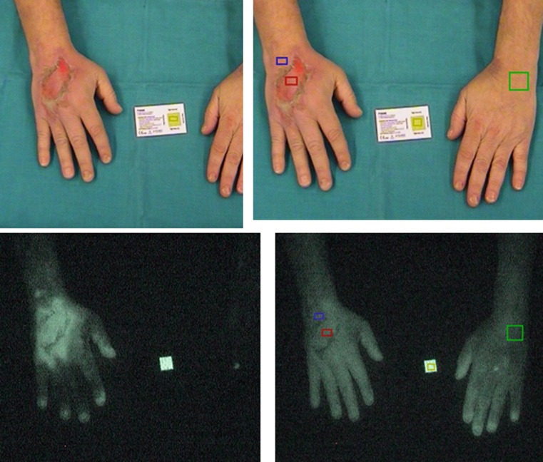
In cancer treatment, extravasation is defined as an inadvertent instillation or leakage of cytotoxic drugs into the perivascular space during infusion. As a dreaded complication of chemotherapy, extravasation has gained increasing attention in recent years. Classified according to their subcutaneous toxicity, three types of cytotoxins have been established: vesicants, irritants and nonvesicant drugs. Vesicant cytotoxic drugs may induce tissue damage, ulceration and tissue necrosis. Although we have established measures to manage extravasation emergencies, prevention is of paramount importance. This may be achieved within hospitals through regular training and education, which is best provided by a specialised and experienced task force including all disciplines involved in cancer therapy. Moreover, clinical and translational studies contribute to a better management of chemotherapy-induced extravasation as shown by our group in recent years. We were able to demonstrate that the evaluation of blood flow by indocyanine green angiography in the extravasation area predicts the extent of damage and the need of future surgical intervention. When a Port-a-Cath® extravasation is detected early, a subcutaneous wash-out procedure was found to be beneficial, corroborated by the analytical evaluation of the removed cytotoxic compound epirubicin. In another study, the tissue distribution of platinum was quantified at the anatomic level in cryosections of various tissues. This novel knowledge complements and supports our current efforts to handle extravasations better. On the other hand, a number of new drugs (chemotherapy, monoclonal antibodies, checkpoint inhibitors etc.) with many open issues to reliably classify their tissue toxicity still require our attention.
Keywords: Chemotherapy complication; Clinical studies; Cytotoxins; Extravasation; Indocyanine green angiography.
Conflict of interest statement
Conflict of interestU. Pluschnig, W. Haslik, R. Bartsch, and R.M. Mader declare that they have no competing interests.
Figures

References
-
- Mader I, Furst-Weger P, Mader RM, Nogler-Semenitz E, Wassertheurer S. Extravasation of cytotoxic agents. 2. Wien: Springer; 2010. - PubMed
Publication types
LinkOut - more resources
Full Text Sources
Other Literature Sources
Research Materials