. 2012 May 14;3(1-3):25-31.
doi: 10.3727/215517912X639496. eCollection 2012 Jan.

Hepatocyte Is a Sole Cell Type Responsible for the Production of Coagulation Factor IX In Vivo

Affiliations

Hepatocyte Is a Sole Cell Type Responsible for the Production of Coagulation Factor IX In Vivo

Kohei Tatsumi et al. Cell Med. .

Abstract

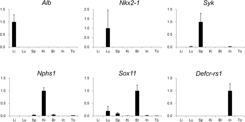
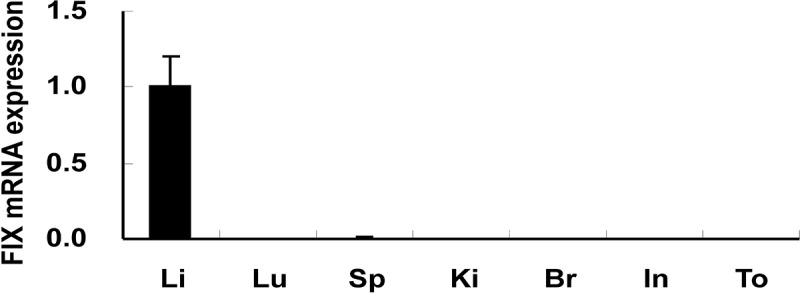
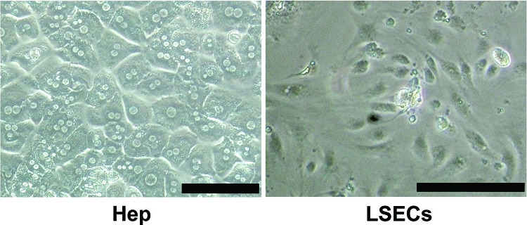
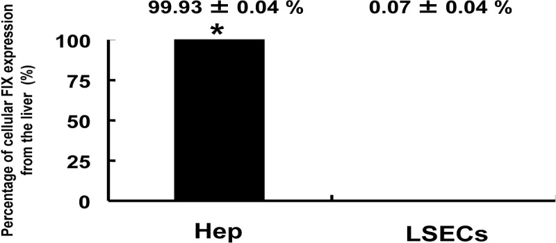
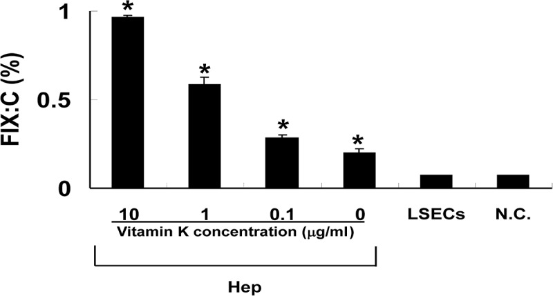
Coagulation factor IX (FIX) is synthesized by hepatocytes, and the lack of this protein causes hemophilia B. Liver nonparenchymal cells, including liver sinusoidal endothelial cells (LSECs) and extrahepatic cells in the body, are scarcely shown to have an ability to synthesize and secrete FIX. The present study investigated the existence of cells responsible for synthesizing FIX other than hepatocytes in mice using gene expression analyses and FIX-specific clotting assays. Among the several organs investigated, including liver, lung, spleen, kidney, brain, intestine, and tongue, FIX mRNA expressions were observed only in the liver. From the liver, hepatocytes and LSECs were isolated. FIX mRNA expression and FIX protein secretion were observed exclusively in the hepatocytes. Furthermore, the clotting activity of FIX secreted from the cultured hepatocytes was found to be dependent on the concentration of vitamin K2. These findings indicated that the hepatocyte is the only cell type that biochemically produces functional FIX in vivo. This highlights the importance of hepatocytes or cells that are fully differentiated toward the hepatic lineage for possible application for regenerative medicine and for targeting gene delivery to establish new cell-based treatments for hemophilia B.

Keywords: Factor IX; Hemophilia B; Hepatocyte; Nonparenchymal cell.


Figures

Figure 1
Figure 1
Validation of the extraction of organ samples. Using extracted organ samples, the gene expression levels of albumin (Alb), NK2 homeobox 1 (Nkx2-1), spleen tyrosine kinase (Syk), nephrosis 1, congenital, Finnish type (nephrin) (Nphs1), sex-determining region Y-box 11 (Sox11), and defensin-related sequence cryptdin peptide (Defcr-rs1) were evaluated by real-time PCR (n=3). Data were normalized by glyceraldehyde-3-phosphate dehydrogenase (GAPDH) expression levels and analyzed by Δ–ΔCt method. Values were graphed as comparative ratios to the levels in their specific organs and were represented as mean ± standard error of the mean (SEM). Li, liver; Lu, lung; Sp, spleen; Ki, kidney; Br, brain; In, Intestine; To, tongue.
Figure 2
Figure 2
Coagulation factor IX (FIX) gene expression in mouse organs. Mouse FIX mRNA expression levels in several mouse organs were determined by real-time PCR (n=4). Data were normalized by glyceraldehyde-3-phosphate dehydrogenase (GAPDH) expression levels and graphed as a comparative ratio to the liver. Values were represented as mean±SEM. Li, liver; Lu, lung; Sp, spleen; Ki, kidney; Br, brain; In, Intestine; To, tongue.
Figure 3
Figure 3
Cell morphology of isolated cell fractions. Liver cells were isolated from the livers of FVB/N mice by a collagenase perfusion method. Hepatocyte (Hep) fraction was purified by Percoll isodensity centrifugation, and liver sinusoidal endothelial cell (LSEC) fraction was condensed by magnetic cell sorting. Isolated cells were seeded on type I collagen-coated six-well culture dishes at a density of 7.5 × 10⁵ cells per well and were cultured for 48 h. Scale bars: 100 μm.
Figure 4
Figure 4
FIX gene expression in fractions of isolated liver cells. Hepatocytes (Hep) and liver sinusoidal endothelial cells (LSECs) were isolated from mouse livers by collagenase perfusion. Mouse FIX mRNA expression levels in each cell fraction were determined by real-time PCR (n=6, respectively). Data were normalized to glyceraldehyde-3-phosphate dehydrogenase (GAPDH) expression levels and graphed as a comparative ratio to the liver. Values were represented as mean±SEM. Asterisks indicate significant differences.
Figure 5
Figure 5
Measurement of FIX activity in the culture medium of cultured liver cells. Isolated hepatocytes and liver sinusoidal endothelial cells were cultured for 16 h in serum-free culture media. Vitamin K2 was added to the media at increasing concentrations from 0.1 to 10 μg/ml. FIX clotting activity in the media was measured by a one-stage clotting assay (n=6, respectively). Data were expressed as the percentage of normal mouse plasma. Values were represented as mean±SEM. Hep, hepatocytes; LSECs, liver sinusoidal endothelial cells; N.C., cell-free control media. Asterisks indicate significant differences versus N.C.

Similar articles

Cited by

References

    1. Berry M. N.; Friend D. S. High-yield preparation of isolated rat liver parenchymal cells: A biochemical and fine structural study. J. Cell Biol. 43:506–520;1969. - PMC - PubMed
    1. Birraux J.; Menzel O.; Wildhaber B.; Jond C.; Nguyen T. H.; Chardot C. A step toward liver gene therapy: Efficient correction of the genetic defect of hepatocytes isolated from a patient with Crigler-Najjar syndrome type 1 with lentiviral vectors. Transplantation 87:1006–1012;2009. - PubMed
    1. Bolton-Maggs P. H.; Pasi K. J. Haemophilias A and B. Lancet 361:1801– 1809; 2003. - PubMed
    1. Boost K. A.; Auth M. K.; Woitaschek D.; Kim H. S.; Hilgard P.; Nadalin S.; Blaheta R. A. Long-term production of major coagulation factors and inhibitors by primary human hepatocytes in vitro: Perspectives for clinical application. Liver Int. 27:832–844;2007. - PubMed
    1. Coutu D. L.; Cuerquis J.; El Ayoubi R.; Forner K. A.; Roy R.; Francois M.; Griffith M.; Lillicrap D.; Yousefi A. M.; Blostein M. D.; Galipeau J. Hierarchical scaffold design for mesenchymal stem cell-based gene therapy of hemophilia B. Biomaterials 32:295–305;2011. - PubMed

LinkOut - more resources